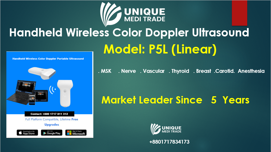
The endorectal handheld wireless color Doppler ultrasound is a versatile and portable diagnostic tool with many applications and benefits in modern medical practice. Below are detailed applications and benefits of this device:
Contact for details:
WhatsApp/Call: 01717811312
Applications
1. Prostate Imaging
- Diagnosis: Identifying prostate enlargement (BPH) or cancer.
- Monitoring: Assessing changes in prostate size or vascularity over time.
2. Rectal Cancer Diagnosis and Staging
- Determining tumor size, invasion depth, and involvement of lymph nodes or surrounding tissues.
3. Evaluation of Pelvic Floor Dysfunction
- Assessing rectal prolapse, fecal incontinence, or muscle weakening.
4. Inflammatory Bowel Disease (IBD)
- Visualizing rectal involvement in Crohn’s disease or ulcerative colitis.
5. Anal Fistula and Abscess Imaging
- Mapping fistula tracts and identifying abscesses to aid surgical planning.
6. Post-Surgical Monitoring
- Ensuring proper healing and detecting complications after rectal or prostate surgeries.
7. Biopsy Guidance
- Assisting in precise needle placement for prostate or rectal biopsies.
8. Radiotherapy Planning
- Providing detailed imaging to guide radiation treatment for rectal or prostate cancer.
9. Sexual Dysfunction Evaluation
- Assessing vascular abnormalities and pelvic blood flow related to erectile dysfunction.
10. Pelvic Tumor Assessment
- Identifying benign or malignant tumors in the rectal or pelvic regions.
11. Hemorrhoids and Vascular Disorders
- Evaluating blood flow in hemorrhoids and rectal varices using Doppler imaging.
12. Rectal Foreign Body Detection
- Localizing foreign objects in the rectum for safe and effective removal.
13. Ischemic Bowel Diagnosis
- Evaluating blood flow to detect ischemia in the rectal area.
14. Endometriosis
- Assessing rectal wall involvement in pelvic endometriosis.
15. Pediatric Anomalies
- Visualizing congenital malformations of the rectum or pelvic region.
16. Rectocele Assessment
- Diagnosing rectocele in women with pelvic organ prolapse.
17. Post-Radiation Monitoring
- Detecting radiation-induced tissue damage or cancer recurrence.
18. Chronic Anorectal Pain
- Identifying structural or vascular causes of persistent rectal pain.
19. Assisted Therapies
- Real-time imaging during treatments like prostate brachytherapy or abscess drainage.
20. Training and Research
- Used in medical training for understanding rectal and pelvic anatomy.
Benefits
1. Portability
- Compact and lightweight, making it ideal for bedside, outpatient clinics, or remote locations.
2. Wireless Connectivity
- Real-time image transfer to smartphones, tablets, or cloud storage for remote interpretation or sharing with specialists.
3. Cost-Effectiveness
- More affordable than traditional cart-based ultrasound machines, lowering diagnostic costs for patients and clinics.
4. High-Quality Imaging
- Provides clear 2D, and Color Doppler images, crucial for evaluating blood flow and detecting abnormalities.
5. Non-Invasive
- Offers a safe and minimally invasive way to evaluate rectal and pelvic organs.
6. Real-Time Imaging
- Enables immediate diagnosis and dynamic evaluation of blood flow and organ movement.
7. Ease of Use
- User-friendly design with intuitive controls, reducing the learning curve for healthcare professionals.
8. Improved Patient Comfort
- Compact probe size minimizes patient discomfort during rectal exams compared to larger devices.
9. Versatility
- Can be used across a wide range of clinical settings, from general practice to specialized oncology and gastroenterology clinics.
10. Enhanced Workflow
- Quick setup and wireless functionality improve efficiency, allowing clinicians to perform more procedures in less time.

