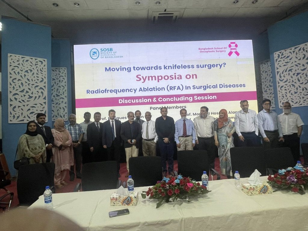
A Milestone in Modern Surgery: Successful Completion of the 1st Symposium on Radiofrequency Ablation (RFA) in Surgical Diseases in Bangladesh
A Milestone in Modern Surgery: Successful Completion of the 1st Symposium on Radiofrequency Ablation (RFA) in Surgical Diseases in Bangladesh
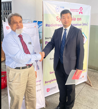
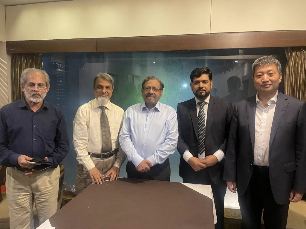
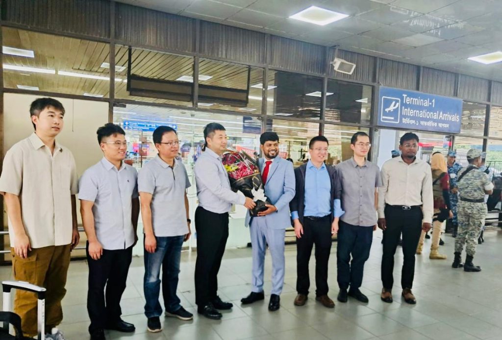
Dhaka, Bangladesh — 16–17 August, 2025 marked a historic moment in the country’s medical landscape as the 1st Symposium on Radiofrequency Ablation (RFA) in Surgical Diseases was successfully held under the banner of the Society of Surgeons of Bangladesh (SOSB). Organized in collaboration with Unique Medi Trade, the Exclusive Distributor of CuraWay Radiofrequency Ablation Systems in Bangladesh, the event brought together national and international experts to share groundbreaking insights on the role of RFA in modern surgery.
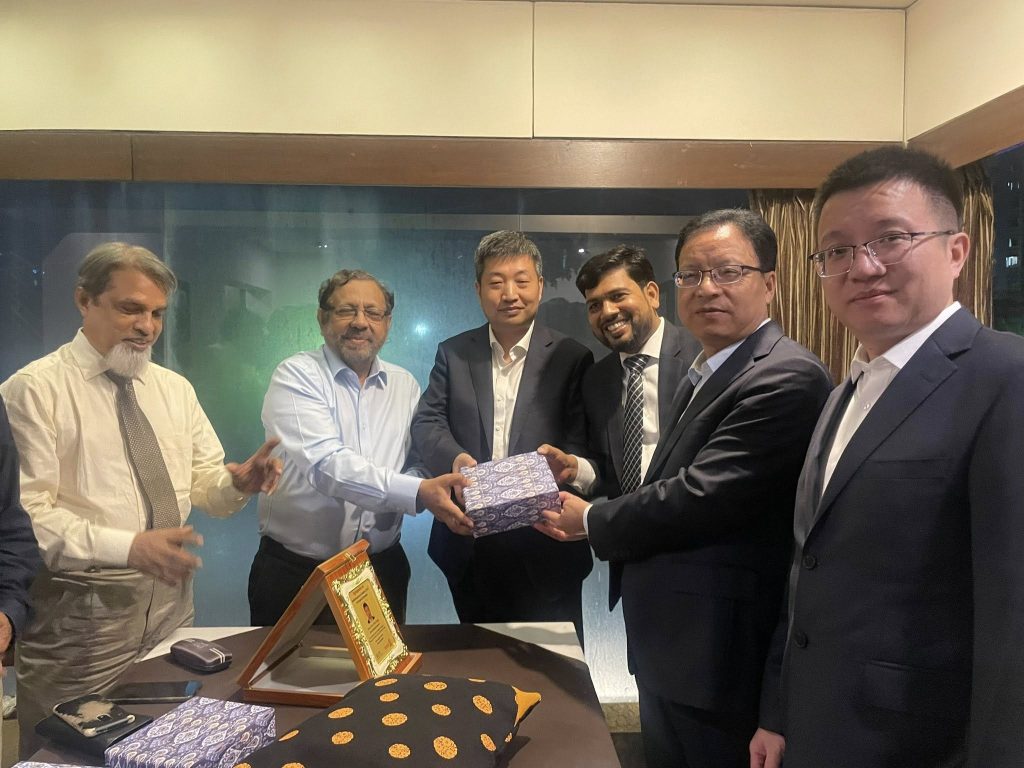
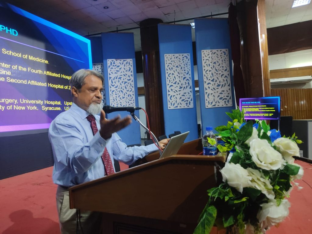
The symposium showcased cutting-edge developments in minimally invasive surgical techniques with a focus on RFA applications in liver tumors, thermal medicine, hemorrhoids, and chronic venous diseases. The presence of distinguished speakers, including Prof. Dr. Tang Zhe from Zhejiang University School of Medicine, China, alongside eminent Bangladeshi experts, added international depth and credibility to the event.
This news feature provides an in-depth look at the symposium, the significance of RFA technology, the insights shared by experts, and the future outlook for surgical innovation in Bangladesh.
What is Radiofrequency Ablation (RFA)?
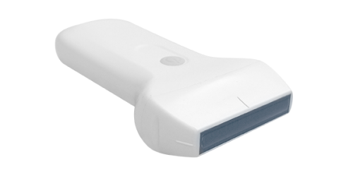
Radiofrequency Ablation is a minimally invasive medical procedure that uses high-frequency alternating current to generate heat and destroy abnormal or diseased tissue. Over the past two decades, RFA has transformed treatment protocols across various specialties, particularly in oncology, gastroenterology, vascular medicine, and pain management.
Globally, RFA is recognized for its safety, precision, and effectiveness. It allows physicians to target tumors, vascular lesions, or other pathological tissues while minimizing damage to surrounding healthy tissue. In many cases, RFA has emerged as an alternative to open surgery, reducing hospital stays, recovery time, and patient discomfort.
For countries like Bangladesh, where the burden of surgical diseases such as liver cancer, venous insufficiency, and colorectal disorders is high, the availability and adoption of RFA technology represent a significant leap toward modernizing healthcare.
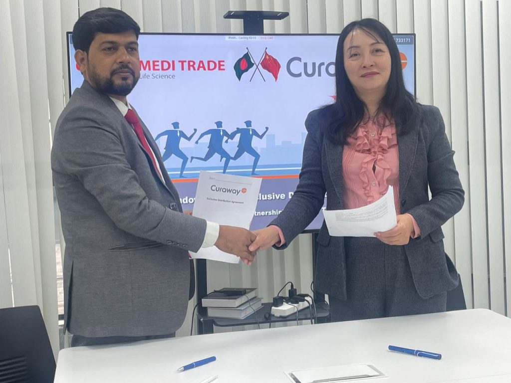
Unique Medi Trade and CuraWay: Bringing Global Innovation to Bangladesh
The successful symposium was made possible through the collaboration of Unique Medi Trade, a leading medical equipment supplier in Bangladesh, and CuraWay, a renowned global manufacturer of Radiofrequency Ablation systems.
CuraWay has established itself as an innovator in advanced surgical technology, with a strong focus on reliability, clinical efficiency, and patient safety. Its RFA systems are widely used in leading hospitals around the world.
Unique Medi Trade, as the exclusive distributor of CuraWay in Bangladesh, has been at the forefront of introducing advanced medical technologies into the country. With its commitment to quality, training, and after-sales support, the company has become a trusted partner for healthcare providers nationwide.
The symposium underscored Unique Medi Trade’s role as not just a distributor, but a pioneer in medical education and clinical transformation. By creating a platform for knowledge exchange, the company demonstrated its commitment to supporting the medical community and advancing surgical practice in Bangladesh.
About the Symposium: A First of Its Kind in Bangladesh
The 1st Symposium on RFA in Surgical Diseases was a milestone event for Bangladesh. Never before had such a focused academic program been arranged in the country on this groundbreaking technology.
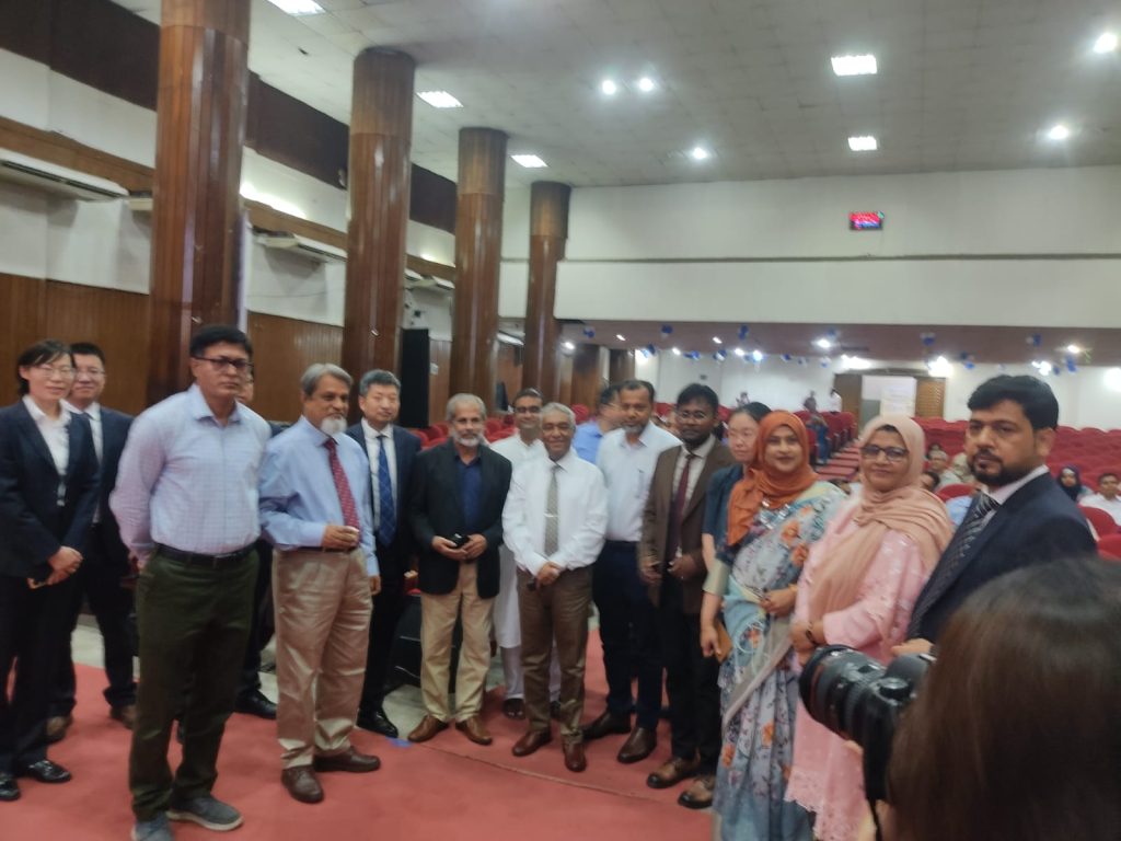
Held on 16–17 August 2025, the symposium provided a rare opportunity for surgeons, oncologists, vascular specialists, and colorectal surgeons to explore the potential of RFA in clinical practice. The program featured four scientific sessions, each addressing a different domain of surgical application.
The participation of an international faculty member, Prof. Dr. Tang Zhe, alongside respected Bangladeshi experts, created a balanced blend of global experience and local relevance. The discussions emphasized practical knowledge, case-based learning, and the future of RFA in Bangladesh’s healthcare system.
Highlights of Scientific Sessions
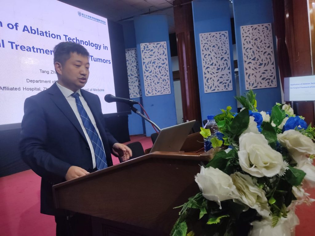
 Application of RFA Ablation Technology in the Surgical Treatment of Liver Tumors
Speaker: Prof. Dr. Tang Zhe, Department of Surgery, The Fourth Affiliated Hospital, Zhejiang University School of Medicine, China.
Prof. Tang Zhe delivered a comprehensive lecture on the role of RFA in managing primary and secondary liver tumors. He highlighted the rising global burden of hepatocellular carcinoma (HCC) and the limitations of traditional surgical resection in many patients.
Key takeaways from his session included:
Minimally Invasive Alternative: RFA provides a curative treatment option for patients who are not candidates for surgery.
Precision in Targeting Tumors: With ultrasound or CT guidance, RFA enables precise ablation of tumor tissue.
Clinical Outcomes: Multiple studies have shown comparable survival rates between RFA and surgical resection in small HCC cases.
Future Directions: Integration of RFA with other modalities such as transarterial chemoembolization (TACE) and immunotherapy could further improve outcomes.
Prof. Tang’s presentation inspired participants by showing how Bangladesh can adopt these protocols to reduce the mortality burden of liver cancer.
 Thermal Medicine in Surgical Diseases: A Brief Overview
Speaker: Prof. M. Mizanur Rahman
Prof. Rahman introduced participants to the broader concept of thermal medicine — the use of heat-based technologies in the management of surgical diseases. His presentation placed RFA within a wider context, including microwave ablation, laser therapy, and high-intensity focused ultrasound (HIFU).
Key highlights:
Thermal medicine offers minimally invasive alternatives for tumors, varicose veins, and benign conditions.
RFA is well-established, with decades of clinical validation.
Bangladesh needs to integrate thermal medicine into its surgical training curriculum to ensure long-term sustainability.
Prof. Rahman emphasized that introducing technologies like RFA is not just about importing machines but about building knowledge ecosystems, where surgeons are trained, patients are educated, and institutions are empowered
.
 Radiofrequency Ablation for Hemorrhoids
Speaker: Dr. Ismat Jahan Lima, Associate Professor, Department of Colorectal Surgery, Shaheed Suhrawardy Medical College; Colorectal & Pelvic Floor Surgeon.
Dr. Lima delivered one of the most engaging sessions of the symposium, focusing on the role of RFA in the treatment of hemorrhoids and anal fistula.
She outlined:
Current Challenges: Conventional hemorrhoidectomy is often associated with significant pain, bleeding, and prolonged recovery.
RFA Advantages: With minimal tissue damage, RFA offers faster recovery, less post-operative pain, and excellent patient satisfaction.
Clinical Evidence: International studies and her own experience demonstrated that RFA is a game-changer in proctology.
Her session resonated strongly with Bangladeshi surgeons, given the high prevalence of hemorrhoidal disease in the country. Dr. Lima’s message was clear: RFA must become part of routine proctology practice in Bangladesh.

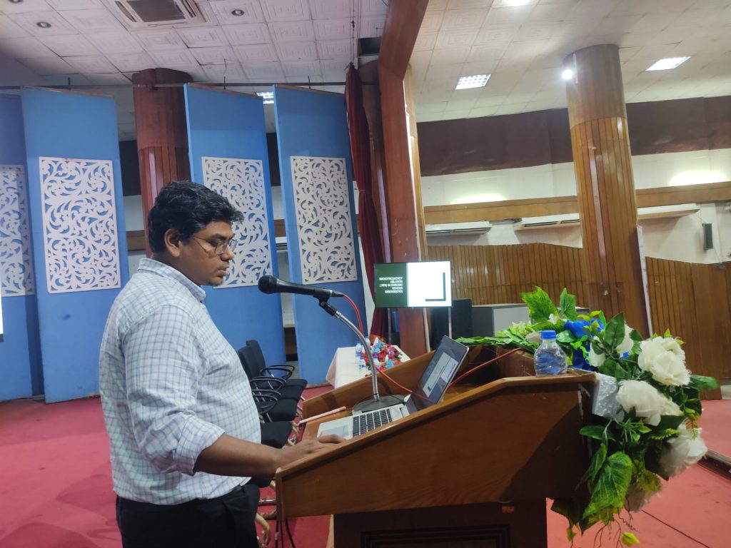
4️⃣ RFA in Chronic Venous Disease
Speaker: Dr. Nur A. Al Amin, Consultant, Endovascular & Vascular Surgeon, IBN SINA Specialized Hospital.
Dr. Al Amin’s lecture focused on the application of RFA in endovenous treatment of varicose veins and chronic venous insufficiency.
Key points:
Endovenous RFA is less invasive than vein stripping or ligation, with better cosmetic results and shorter recovery.
Patients treated with RFA often return to work within days, compared to weeks with traditional surgery.
Complication rates are low, and long-term recurrence is minimal.
His talk emphasized how vascular surgery in Bangladesh can benefit tremendously from adopting RFA, given the large patient population suffering from varicose veins.
Impact on Bangladesh’s Healthcare System
The symposium highlighted the transformative potential of RFA technology in Bangladesh:
Improved Patient Outcomes: Less invasive, safer, and faster recovery.
Reduced Healthcare Burden: Shorter hospital stays and quicker return to normal life.
Skill Development: Training opportunities for local surgeons to adopt cutting-edge techniques.
Healthcare Modernization: Positioning Bangladesh as a regional leader in advanced surgical care.
Unique Medi Trade’s role was critical. By bridging the gap between global innovation and local clinical practice, the company is helping Bangladesh’s healthcare system keep pace with international standards.
Future Outlook: The Road Ahead for RFA in Bangladesh
The successful completion of this symposium is only the beginning. Going forward:
Training Programs: Regular workshops and hands-on sessions will be required to ensure more surgeons are skilled in RFA.
Hospital Adoption: Public and private hospitals need to incorporate RFA into their surgical departments.
Research & Data Collection: Local studies will help validate RFA outcomes in the Bangladeshi population.
Patient Awareness: Educating patients about minimally invasive options will increase acceptance and demand.
Unique Medi Trade has already announced plans to continue collaborating with SOSB and international experts to strengthen clinical training, provide equipment support, and expand RFA services nationwide.
Closing Remarks
The 1st Symposium on Radiofrequency Ablation (RFA) in Surgical Diseases in Bangladesh was more than just an academic program — it was a landmark in the modernization of surgical practice in the country.
With the support of CuraWay and the leadership of Unique Medi Trade, surgeons in Bangladesh are now better equipped to embrace minimally invasive, patient-centered care.
As Prof. Tang Zhe highlighted during his session, “The future of surgery lies in innovation that makes treatment safer, faster, and more effective. Radiofrequency Ablation is one of the most important steps in this direction.”
Unique Medi Trade’s efforts in hosting this symposium reflect its commitment to empowering doctors, advancing healthcare, and improving patient outcomes in Bangladesh.
This historic event will be remembered as the starting point of a new era of RFA-based surgical excellence in the country.