Search

Digital Bluetooth Wireless Smartphone Stethoscope

The Ultimate Guide to Digital Bluetooth Wireless Stethoscopes

The Ultimate Guide to Digital Bluetooth Wireless Stethoscopes

Revolutionizing Auscultation for the Modern Healthcare Era

1. Introduction

The stethoscope has long been the iconic symbol of the medical profession. From the days of René Laennec’s wooden monaural invention in 1816 to the dual-head Littmann of the 20th century, the humble stethoscope has seen only incremental changes — until now.

Welcome to the digital era, where Digital Bluetooth Wireless Stethoscopes are revolutionizing the way clinicians listen, diagnose, and treat. These devices are more than just futuristic accessories — they’re powerful diagnostic tools designed to meet the complex demands of modern medicine.

Whether you're a physician, nurse, medical student, or remote healthcare provider, this guide will walk you through everything you need to know about digital Bluetooth stethoscopes — from how they work, to how to choose one, and what the future holds.

Digital Bluetooth Wireless Smartphone Stethoscope

Buy Now

2. Evolution of the Stethoscope


2.1 The Invention

The original stethoscope was developed by French physician René Laennec in 1816, using a wooden tube to amplify chest sounds.

2.2 From Acoustic to Amplified

Over the decades, innovations led to binaural stethoscopes, dual diaphragms, and acoustic enhancements. In the late 20th century, companies like 3M Littmann introduced electronic amplification.

2.3 Enter the Digital Age

The 21st century introduced:

  • Sound amplification up to 40x
  • Noise reduction algorithms
  • Bluetooth connectivity
  • Mobile app integration
  • Telemedicine compatibility

Today’s stethoscopes are smart, connected, and designed for real-time diagnostics and data sharing.


3. What is a Digital Bluetooth Wireless Stethoscope?

A Digital Bluetooth Wireless Stethoscope is an advanced diagnostic tool that:

  • Converts acoustic body sounds (like heartbeats, breath sounds) into digital signals
  • Allows amplification, visualization, and recording
  • Uses Bluetooth to connect with smartphones, tablets, or computers
  • Enables wireless auscultation and remote sharing

It’s not just a better stethoscope. It’s a complete digital auscultation system.


4. How Does It Work?

Digital stethoscopes combine hardware and software components:

4.1 Sensor (Chest Piece)

  • Picks up vibrations from the body (e.g., heart or lung sounds)
  • Transduces them into electronic signals

4.2 Digital Signal Processor (DSP)

  • Filters, amplifies, and processes the signals
  • Eliminates ambient noise using advanced algorithms

4.3 Bluetooth Module

  • Sends the processed audio to paired devices (headphones, phones, computers)

4.4 Mobile App

  • Displays sound waveforms in real time
  • Records audio for playback or sharing
  • Offers cloud backup, patient profiles, and AI insights (in some models)

5. Key Features and Technology

Let’s break down the cutting-edge tech behind these devices:

 
Feature Description
Amplification Up to 40x normal auscultation
Noise Cancellation Digital filters remove ambient interference
Bluetooth Connectivity Seamless pairing with smart devices
Recording & Playback Store, replay, or forward sounds to specialists
Cloud Storage Patient data saved securely online
Visualization Waveform display for more precise diagnostics
AI Integration Some models analyze sound patterns to detect anomalies

6. Benefits Over Traditional Stethoscopes

6.1 Clinical Accuracy

Amplification and filtering improve detection of faint murmurs and subtle pulmonary abnormalities.

6.2 Telemedicine

Remote auscultation becomes possible — essential during pandemics and for rural healthcare.

6.3 Documentation & Education

Recorded sounds enhance patient records and support medical training.

6.4 Accessibility

Hearing-impaired clinicians can use visual feedback or connect to hearing aids.

6.5 Integration

Easily link patient data with Electronic Medical Records (EMRs).


7. Use Cases and Applications

7.1 General Practice

Routine physicals with enhanced precision.

7.2 Cardiology

Clearer detection of systolic/diastolic murmurs, arrhythmias, and more.

7.3 Pulmonology

Auscultate wheezing, rales, crackles — even remotely.

7.4 Pediatrics

Capture and record fast-changing heart/lung sounds in children.

7.5 Medical Education

Replay and analyze audio for students during rounds or lectures.

7.6 Home & Telehealth Monitoring

Used by visiting nurses or even patients at home under doctor guidance.

8. Top Digital Stethoscope Brands

Several manufacturers are at the forefront of digital stethoscope innovation. Here are some of the most reputable:

HMYL Digital Stethoscope

  • Wireless, smartphone-friendly, and designed for modern clinicians.
  • Features: Bluetooth audio transmission, sound recording, lightweight build.

9. Buyer’s Guide: Choosing the Right Device

When selecting the right digital stethoscope, consider:

✔️ Your Medical Specialty

  • Cardiology: Go for devices with ECG and highest sound fidelity.
  • Primary Care: A mid-range model with recording and Bluetooth is usually enough.
  • Telehealth: Must include app support and wireless sharing.

✔️ Audio Quality

  • Look for amplification power, frequency range, and noise reduction.

✔️ Battery Life

  • Some last up to 60 hours on a single charge. Others require frequent recharging.

✔️ Compatibility

  • Works with iOS, Android, and optionally with EMR software.

✔️ Storage & Sharing

  • Consider models that allow audio recording, cloud storage, and waveform sharing.

✔️ Price Range

  • Entry-level: $100–$200
  • Mid-range: $250–$350
  • High-end: $400–$600+

10. Clinical Validation and Certifications

Any medical diagnostic device should meet regulatory requirements. Check for:

CE Mark

  • Ensures compliance with European health, safety, and environmental protection standards.

ISO Certification

  • Verifies quality control processes in manufacturing.

Without proper certifications, avoid using the device in professional settings.


11. Integration with Mobile Devices

📱 Smartphone Integration

  • Connects via Bluetooth to a dedicated app.
  • Shows records sounds, and even offers patient tagging.

12. Data Privacy and Security

Because these devices collect and transmit patient data:

  • Use devices with end-to-end encryption.
  • Confirm whether local or cloud storage is used.
  • Always keep apps and firmware updated.

13. Troubleshooting and Maintenance Tips

To get the best performance and longevity out of your digital stethoscope, follow these best practices:

🔧 Troubleshooting Common Issues

  • Bluetooth Not Connecting
    → Ensure device is charged and Bluetooth is enabled on your phone. Reboot both devices.
  • Poor Sound Quality
    → Check for firmware updates, adjust volume on the app, and make sure the diaphragm is clean.
  • App Crashing or Freezing
    → Close background apps, clear cache, and check for software updates.
  • Recording Not Saving
    → Verify storage permissions for the app and check available memory.

🧼 Cleaning and Care

  • Use alcohol wipes to clean the diaphragm and tubing (if present).
  • Avoid submerging in liquid.
  • Do not use abrasive cleaners.
  • Store in a cool, dry place in its case.

14. Common Myths and Misconceptions

Let’s bust some myths:

“Digital stethoscopes are just expensive toys.”

✅ Truth: They are powerful diagnostic tools with amplified audio, recording, and sharing capabilities that improve patient care.

“They’re too complicated to use.”

✅ Truth: Most models are intuitive, with one-button operation and user-friendly apps.

“Bluetooth means low sound quality.”

✅ Truth: Modern Bluetooth technology ensures high-fidelity audio, often better than acoustic auscultation.

“They can’t be used for real diagnoses.”

✅ Truth: Many devices are FDA-approved and used in hospitals, clinics, and universities worldwide.


15. Price Range and Budget Considerations

Here’s a general guide to pricing:

 
Budget Features Examples
Under $150 Basic Bluetooth stethoscopes, no recording Entry-level
$150–$300 Noise reduction, sound recording, app support HMYL Premium
$300–$600 Advanced audio, AI integration, ECG Eko DUO, Littmann CORE
$600+ Specialty devices, multi-sensor tech, hospital-grade Thinklabs One Pro, custom telemedicine kits

16. Global Market Trends

The demand for digital stethoscopes is rising rapidly. Here's why:

🌍 Telemedicine Growth

Driven by the pandemic and rural healthcare expansion, remote auscultation is becoming a norm.

📈 Market Forecast

The global digital stethoscope market is projected to exceed USD 1.5 billion by 2030, with CAGR of 6–9%.

💼 Healthcare Digitization

With EMRs and AI becoming essential, smart stethoscopes are a critical part of the diagnostic ecosystem.

🔬 AI Diagnostics

AI-assisted auscultation is evolving. Devices now suggest murmurs, wheezing, or arrhythmia detection.


17. Future of Digital Auscultation

Here’s what’s on the horizon:

🤖 AI Integration

AI will not just detect anomalies — it’ll predict cardiac events, recommend treatment, and even generate reports.

☁️ Cloud-First Ecosystems

Doctors will collaborate across continents by listening to the same chest sound in real time.

📲 All-in-One Diagnostic Kits

Integrated with ECG, BP, SpO2, and even ultrasound into pocket-sized telehealth kits.

🧠 Augmented Auscultation

Combine auscultation data with imaging and lab results for deeper diagnostic insights.


18. Frequently Asked Questions (FAQs)

Q: Can I use a digital stethoscope for pediatrics?

A: Yes, many models come with pediatric and neonatal modes for high-frequency lung and heart sounds.

Q: Is Bluetooth safe for medical use?

A: Absolutely. Medical-grade Bluetooth operates within secure, encrypted standards.

Q: Can I share recordings with other doctors?

A: Yes. Most apps allow secure sharing via email, cloud, or even live streaming.

Q: How long does the battery last?

A: Most digital stethoscopes last 8–60 hours per charge, depending on the model and usage.

Q: Are digital stethoscopes approved by medical boards?

A: Many are FDA-cleared and CE-certified. Always verify before purchasing.


19. Final Thoughts

Digital Bluetooth wireless stethoscopes are not just a novelty — they’re essential tools in a rapidly evolving medical landscape.

Whether you’re diagnosing heart murmurs, listening to lung crackles, teaching students, or consulting via telehealth, a smart stethoscope enhances your diagnostic capabilities.

🛒 Looking to buy one?
Check out local suppliers like Unique Medi Trade in Bangladesh or authorized dealers near you. Always prioritize certified models with after-sales support.


Remember: Medicine is changing. Your tools should too.

📲 Ready to go digital? Embrace the stethoscope of the future — because your patients deserve the best.

Conclusion

Digital Bluetooth Wireless Stethoscopes are more than just modern gadgets—they’re vital tools in reshaping how healthcare is delivered globally. With their powerful capabilities and expanding accessibility, they bridge gaps in diagnostics, training, and care coordination. Whether you're a practicing clinician, a medical educator, or a tech-savvy student, these devices are your gateway to smarter, faster, and more connected healthcare.
radiofrequency-ablation-of-haemorrhoids-fistula-under-local-anaesthesia

Radiofrequency Ablation (RFA) vs. Laser Treatment for Hemorrhoid & Anal Fistula: A Comparative Analysis

Radiofrequency Ablation (RFA) vs. Laser Treatment for Hemorrhoid & Anal Fistula: A Comparative Analysis. When choosing a minimally invasive procedure for hemorrhoid and anal fistula treatment, both Radiofrequency Ablation (RFA) and Laser treatment are widely used. However, RFA is emerging as a superior alternative due to its enhanced precision, cost-effectiveness, and better post-operative outcomes.


Key Comparisons: RFA vs. Laser

Feature Radiofrequency Ablation (RFA) Laser Treatment
Tissue Damage Minimal, controlled ablation More thermal spread, potential damage to surrounding tissue
Post-Operative Pain Less pain due to precise energy delivery Higher due to thermal effect on surrounding tissues
Healing & Recovery Time Faster, with quicker wound healing Slower due to increased tissue inflammation
Procedure Cost More cost-effective Higher equipment and procedure cost
Recurrence Rate Lower recurrence due to deep coagulation May have a higher recurrence risk in complex cases
Equipment Maintenance Easy to maintain and more affordable Expensive maintenance and laser fiber replacement

Why Surgeons Prefer RFA Over Laser?

Better Patient Comfort: RFA ensures less pain and faster recovery.
More Precision & Safety: Controlled energy minimizes the risk of tissue damage.
Lower Cost, Higher Efficiency: RFA is more cost-effective than laser with similar or better outcomes.
Long-Term Success: Studies show lower recurrence rates in RFA-treated cases.

Conclusion

While both techniques are effective, RFA offers a safer, more affordable, and patient-friendly solution for hemorrhoid and anal fistula treatment.

🚀 Upgrade to RFA for superior surgical outcomes!
📞 Contact us today to learn more. +8801717811312
📍 Unique Medi Trade

Bone Marrow Biopsy Needle: Uses, Types, and Procedure

Understanding Bone Marrow Biopsy Needle: Uses, Types, and Procedure

Introduction

A bone marrow biopsy needle is a specialized medical instrument used to extract a sample of bone marrow tissue for diagnostic purposes. Bone marrow is the soft, spongy tissue found inside bones that produces blood cells. A biopsy of this tissue is often necessary to diagnose various hematological diseases, including leukemia, lymphoma, anemia, and other bone marrow disorders. The biopsy procedure requires a precise and well-designed needle to ensure minimal discomfort and an adequate sample for analysis.

This blog post provides an in-depth look at bone marrow biopsy needles, their types, uses, and the procedure involved in obtaining a bone marrow sample.

What is a Bone Marrow Biopsy Needle?

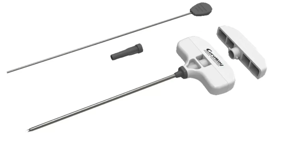
A bone marrow biopsy needle is a hollow, cylindrical medical device designed for extracting a sample of bone marrow tissue. It is made from high-quality stainless steel and comes with ergonomic handles for better control. The needle has a sharp, beveled tip to penetrate the bone effectively while minimizing trauma to surrounding tissues.

Uses of Bone Marrow Biopsy Needle

Bone marrow biopsy needles are primarily used for:

  1. Diagnosis of Blood Disorders: These include leukemia, lymphoma, multiple myeloma, and myelodysplastic syndromes.
  2. Evaluation of Anemia: In cases where the cause of anemia is unclear, a bone marrow biopsy may be performed.
  3. Monitoring Cancer Treatment: Biopsies help assess how well treatments such as chemotherapy are working.
  4. Investigating Bone Marrow Infections: To identify infections that may not be detectable through blood tests.
  5. Stem Cell Analysis: To evaluate the bone marrow before a stem cell transplant.

Components of a Bone Marrow Biopsy Needle

A typical bone marrow biopsy needle consists of the following parts:

  1. Cannula: The hollow tube through which the sample is collected.
  2. Stylet: A sharp inner rod that helps pierce the bone.
  3. Hub: The handle or grip of the needle, allowing precise control.
  4. Depth Markings: Many needles have measurement markings to ensure accurate penetration depth.
  5. Specimen Retrieval Notch: A feature on some needles that aids in sample collection.

Bone Marrow Biopsy Procedure

Preparation

Before performing a bone marrow biopsy, the patient undergoes certain preparations:

  • The procedure is explained to the patient.
  • Local anesthesia is applied to numb the area.
  • The patient is positioned either on their side or stomach, depending on the biopsy site.

Step-by-Step Procedure

  1. Selection of Biopsy Site:
    • The posterior iliac crest (hip bone) is the most common site.
    • In some cases, the sternum (breastbone) or tibia (shin bone) may be used.
  2. Insertion of the Needle:
    • A small incision is made in the skin.
    • The biopsy needle is carefully inserted into the bone with a twisting motion.
    • Once the needle reaches the marrow cavity, the stylet is removed.
  3. Sample Collection:
    • The physician applies gentle pressure to extract the marrow sample.
    • The sample is removed carefully to maintain its integrity.
  4. Needle Removal and Dressing:
    • The needle is withdrawn slowly.
    • A sterile bandage is applied to the biopsy site.

Post-Procedure Care and Recovery

After the biopsy, patients may experience slight discomfort or bruising. The following care measures are recommended:

  • Avoid strenuous activity for at least 24 hours.
  • Keep the biopsy site clean and dry.
  • Over-the-counter pain relievers may be taken if needed.
  • Patients should monitor for signs of infection such as swelling, redness, or excessive pain.

Potential Risks and Complications

While a bone marrow biopsy is a relatively safe procedure, there are some potential risks:

  • Pain and Discomfort: Mild to moderate pain at the biopsy site is common.
  • Bleeding: Patients with bleeding disorders may experience prolonged bleeding.
  • Infection: Though rare, infections can occur if proper sterilization techniques are not followed.
  • Bruising or Hematoma: Minor bruising is expected but usually resolves within a few days.

Choosing the Right Bone Marrow Biopsy Needle

Selecting the appropriate needle depends on several factors:

  • Patient Age and Condition: Pediatric patients may require smaller needles.
  • Procedure Type: Some needles are designed for aspiration, while others are for biopsy.
  • Physician’s Preference: Different doctors may have preferences based on ease of use and efficiency.

Advancements in Bone Marrow Biopsy Needles

Modern innovations have led to improved biopsy needles with enhanced features, such as:

  • Ergonomic Designs: Better grip and control reduce procedural fatigue.
  • Single-Use Needles: Minimize the risk of cross-contamination.
  • Enhanced Tip Designs: Provide smoother penetration and less patient discomfort.
  • Vacuum-Assisted Collection: Some newer models help improve sample retrieval.

Conclusion

A bone marrow biopsy needle is an essential tool in diagnosing and monitoring various blood-related disorders. With advancements in design and technology, these needles have become more efficient and less painful for patients. Physicians must choose the appropriate needle based on the procedure type, patient needs, and desired sample quality. Understanding the role of biopsy needles in medical diagnostics highlights their significance in improving patient outcomes and advancing hematological research.

By using high-quality, well-designed biopsy needles, healthcare professionals can ensure accurate diagnoses and better patient experiences. If you have any concerns about a bone marrow biopsy, consult with a healthcare provider for guidance and reassurance.

radiofrequency-ablation-of-haemorrhoids-fistula-under-local-anaesthesia

Working Principle of Radiofrequency Ablation (RFA) for Surgery

Working Principle of Radiofrequency Ablation (RFA) for tumors, Hemorrhoids & Anal Fistule , Nerve ablation (pain management), and Varicose veins Surgery 

Radiofrequency Ablation (RFA) is a minimally invasive procedure that uses high-frequency alternating current (350–500 kHz) to generate controlled heat and destroy targeted tissues. It is widely used in tumors, hemorrhoids, anal fistulas, nerve ablation (pain management), and varicose veins surgery.

📞 Contact

Unique Medi Trade

📞 +8801717811312 📧 Email: uniquemeditrade21@gmail.com


The RFA process follows three key mechanisms:

  1. Ionic Agitation → Heat Generation
  2. Protein Denaturation → Cell Death
  3. Tissue Collapse → Shrinkage or Absorption

1. Ionic Agitation – Heat Generation

  • When RF energy is applied through an electrode, it creates a rapidly alternating electric field in the tissue.
  • Charged ions (e.g., Na⁺, K⁺, Cl⁻) within the extracellular and intracellular fluid begin to vibrate and oscillate due to this high-frequency field.
  • This rapid movement of ions causes friction, which generates heat inside the tissue.
  • The temperature rises (typically 60–100°C), initiating thermal damage.

🟢 Effect: Produces controlled heating within the target tissue without affecting surrounding structures significantly.


2. Protein Denaturation – Cellular Destruction

  • As the temperature increases beyond 60°C, proteins within the cell begin to denature (lose their natural structure).
  • This leads to:
    Enzyme inactivation → Disrupts cellular metabolism.
    Membrane disruption → Cell walls lose integrity.
    Coagulative necrosis → The tissue becomes non-functional and starts breaking down.

🟢 Effect: The targeted cells lose function and die, preventing abnormal tissue from growing or spreading.


3. Tissue Collapse – Structural Breakdown

  • After the proteins denature, the affected tissue shrinks and dehydrates, leading to:
    Closure of blood vessels → Stops further blood supply (used in vein ablation).
    Fibrotic scar formation → The body absorbs dead tissue over time.
    Loss of abnormal function → Effective for tumors, Hemorrhoids & Anal Fistule , nerve ablation (pain management), and varicose veins.

🟢 Effect: The damaged tissue collapses, shrinks, or gets resorbed, completing the ablation process.


1. RFA for Tumors (Liver, Kidney, Lung, Bone, Thyroid, etc.)

Working Mechanism:

  • A thin probe (electrode) is inserted into the tumor under ultrasound, CT, or MRI guidance.
  • Radiofrequency energy heats the tumor to 60–100°C, causing coagulative necrosis.
  • The tumor cells die, and the body gradually absorbs or scars off the tissue over time.

Effective for: Liver cancer, kidney cancer, lung nodules, bone tumors, thyroid nodules.
Advantage: Minimally invasive, quick recovery, and can be repeated if needed.


2. RFA for Hemorrhoids & Anal Fistulas

Working Mechanism:

  • A needle-like RFA probe is inserted into the hemorrhoidal tissue or fistula tract.
  • The RF current generates heat, leading to vascular coagulation and tissue shrinkage.
  • For hemorrhoids, the blood supply is cut off, and the swollen veins shrink and scar off.
  • For anal fistulas, the abnormal tract is ablated and sealed to promote healing.

Effective for: Grade 2–4 hemorrhoids, complex anal fistulas.
Advantage: Less bleeding, minimal post-operative pain, faster healing compared to traditional surgery.


3. RFA for Nerve Ablation (Pain Management)

Working Mechanism:

  • A thin RF catheter is placed near the target nerve under fluoroscopy (X-ray) or ultrasound guidance.
  • The RF energy disrupts the pain-conducting nerve fibers, preventing pain signals from reaching the brain.
  • This provides long-lasting pain relief without the need for medication.

Effective for: Chronic back pain, arthritis, trigeminal neuralgia, sciatica, post-surgical pain.
Advantage: Non-surgical, precise targeting, relief lasts from months to years.


4. RFA for Varicose Veins Surgery (Endovenous RFA Ablation)

Working Mechanism:

  • A thin RFA catheter is inserted into the affected vein under ultrasound guidance.
  • The RF energy heats the vein walls, causing collagen contraction and vein closure.
  • The body reroutes blood flow to healthier veins, and the closed vein gradually disappears.

Effective for: Large varicose veins, chronic venous insufficiency (CVI).
Advantage: Minimally invasive, no surgical scars, faster recovery than vein stripping.


Conclusion

Radiofrequency Ablation (RFA) is a highly effective, safe, and minimally invasive technique for various conditions:

  • Tumors → Destroys cancer cells with heat.
  • Hemorrhoids & Fistulas → Shrinks and seals abnormal tissues.
  • Nerve Ablation → Blocks pain signals for long-term relief.
  • Varicose Veins → Closes diseased veins and redirects blood flow.
 

📞 Contact

Unique Medi Trade

📞 +8801717811312 📧 Email: uniquemeditrade21@gmail.com

Back to Top
Product has been added to your cart