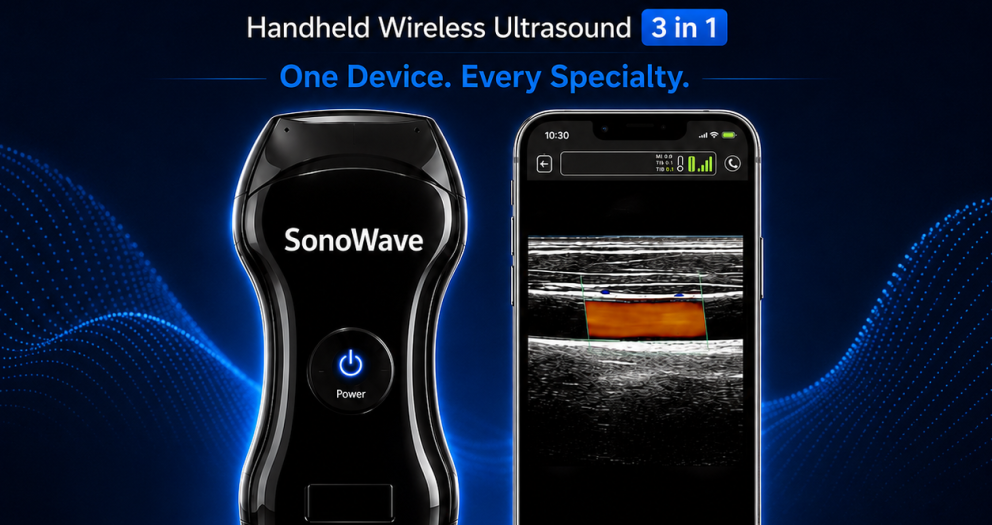
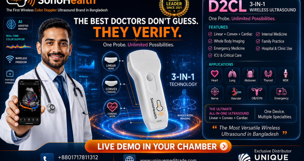
The Ultimate 3-in-1 SonoHealth Handheld Wireless Color Doppler Ultrasound for Modern Medicine
1. Introduction
The Evolution of Modern Diagnostic Imaging
Over the past century, medical imaging has fundamentally transformed the practice of medicine. From the discovery of X-rays in 1895 to the development of computed tomography (CT), magnetic resonance imaging (MRI), and modern ultrasound systems, diagnostic technology has continually expanded physicians' ability to visualize the human body without invasive procedures. Today, medical imaging is no longer confined to radiology departments; it has become an integral part of everyday clinical decision-making across virtually every medical specialty.
Among these innovations,
ultrasound has emerged as one of the most versatile, safe, and accessible diagnostic tools in healthcare. Unlike ionizing imaging modalities, ultrasound provides
real-time visualization, is non-invasive, radiation-free, repeatable, and capable of delivering immediate clinical information at the patient's bedside. These characteristics have made ultrasound indispensable in emergency medicine, intensive care, cardiology, internal medicine, surgery, anesthesiology, obstetrics, vascular medicine, musculoskeletal imaging, and many other disciplines.
The healthcare landscape is also changing rapidly. Modern physicians are expected to diagnose faster, make evidence-based decisions, reduce unnecessary investigations, and improve patient outcomes while operating in increasingly busy clinical environments. These demands have accelerated the adoption of
Point-of-Care Ultrasound (POCUS)—a model in which ultrasound becomes an extension of the clinician's physical examination rather than a separate diagnostic procedure.
Why Ultrasound Has Become the New "Clinical Stethoscope"
For more than two centuries, the stethoscope has symbolized the medical profession. It remains an invaluable instrument for listening to heart, lung, and vascular sounds. However, while auscultation provides indirect clues about a patient's condition, it cannot visualize anatomy or pathology.
Modern ultrasound fills this gap by allowing clinicians to
see what they previously could only infer.
This evolution has led many experts to describe handheld ultrasound as
"the new clinical stethoscope." The comparison is not intended to replace the traditional stethoscope but to emphasize the growing role of bedside imaging in contemporary medicine. With handheld ultrasound, physicians can evaluate cardiac function, detect pleural effusion, assess lung pathology, identify free abdominal fluid, guide vascular access, examine superficial structures, and perform focused assessments within minutes—all during the initial patient encounter.
The philosophy behind this transformation can be summarized in one powerful statement:
There is a huge difference between what you think and what you can see.
By providing immediate visualization, ultrasound reduces diagnostic uncertainty, supports earlier interventions, and enhances clinical confidence. In emergency departments, intensive care units, outpatient clinics, operating rooms, ambulances, and even remote healthcare settings, handheld ultrasound is rapidly becoming an essential extension of the physical examination.
Introducing the SonoHealth D2CL
The
SonoHealth D2CL represents the next generation of handheld Point-of-Care Ultrasound technology. Designed to meet the evolving needs of modern physicians, it combines
Linear, Convex, and Cardiac imaging capabilities into a single wireless device, eliminating the need to carry multiple probes for different examinations.
Unlike conventional cart-based ultrasound systems that are often restricted to dedicated imaging departments, the D2CL is engineered for mobility, flexibility, and immediate bedside use. Its compact design, wireless connectivity, and compatibility with
iOS, Android, and Windows platforms enable clinicians to perform high-quality ultrasound examinations virtually anywhere patient care is delivered.
The D2CL supports advanced imaging modes, including
B-mode, Color Doppler, Power Doppler Imaging (PDI), and Pulse Wave Doppler (PW), allowing physicians to evaluate anatomical structures, blood flow, and cardiac function in real time. Whether assessing a trauma patient in the emergency department, performing focused cardiac ultrasound in the ICU, guiding vascular access during anesthesia, or examining abdominal pathology in an outpatient clinic, the D2CL is designed to provide rapid, reliable imaging that integrates seamlessly into everyday clinical workflow.
Its
3-in-1 probe architecture distinguishes it from many handheld systems by enabling clinicians to switch between superficial, abdominal, and cardiac imaging without changing devices. This versatility supports a wide range of medical specialties and makes the D2CL a practical solution for hospitals, clinics, medical colleges, ambulance services, rural healthcare programs, and telemedicine initiatives.
Overview of This Review
This comprehensive review explores the SonoHealth D2CL from both a clinical and practical perspective. Rather than focusing solely on technical specifications, the article examines how the device can contribute to modern patient care across diverse healthcare settings.
The review discusses the evolution of Point-of-Care Ultrasound, the advantages of wireless imaging, the engineering behind the D2CL, its imaging performance, and its applications in emergency medicine, critical care, cardiology, internal medicine, surgery, anesthesiology, vascular medicine, musculoskeletal imaging, and medical education. It also evaluates workflow integration, digital connectivity, and the potential benefits for hospitals and healthcare institutions adopting portable ultrasound technology.
By the end of this review, readers will gain a clear understanding of the clinical capabilities of the SonoHealth D2CL, the role of handheld ultrasound in contemporary medicine, and the factors healthcare professionals should consider when selecting a Point-of-Care Ultrasound system for their practice.
As medicine continues to shift toward faster, more evidence-based, and patient-centered care, portable ultrasound is becoming more than a diagnostic device—it is becoming an essential clinical companion. The SonoHealth D2CL has been developed to support this transformation by bringing advanced ultrasound imaging directly into the hands of physicians, wherever patient care is needed.
2. The Evolution of Point-of-Care Ultrasound (POCUS)
From the Radiology Department to the Patient's Bedside
The practice of medicine has always been driven by one fundamental objective: obtaining accurate clinical information as quickly and safely as possible. Throughout history, physicians have relied on physical examination, clinical experience, and diagnostic investigations to understand disease. However, one of the most transformative advances in modern medicine has been the evolution of
Point-of-Care Ultrasound (POCUS)—a technology that allows clinicians to visualize anatomy and physiology in real time, exactly where patient care is taking place.
Today, POCUS is no longer considered merely an imaging tool. It has become an extension of the physician's clinical examination, providing immediate answers to focused clinical questions and enabling faster, evidence-based decision-making across almost every medical specialty.
The History of Ultrasound
Medical ultrasound has a rich history spanning more than seven decades. The underlying principles of ultrasound originated from sonar technology developed during the early twentieth century. Following World War II, researchers began adapting high-frequency sound waves for medical imaging, leading to the first diagnostic ultrasound systems in the 1950s.
Early ultrasound machines were large, technically complex, and produced relatively low-resolution images. Their primary applications were in obstetrics, where they revolutionized fetal assessment by providing a safe, radiation-free method for monitoring pregnancy.
As technology advanced, ultrasound expanded into numerous specialties, including cardiology, abdominal imaging, vascular medicine, urology, musculoskeletal imaging, and emergency medicine. Improvements in transducer design, digital signal processing, Doppler technology, and image resolution transformed ultrasound into one of the most versatile diagnostic modalities available.
For many years, however, ultrasound remained confined to radiology departments and specialized imaging centers. Large cart-based systems required dedicated examination rooms, trained sonographers, and scheduled appointments, limiting immediate access for frontline clinicians.
The Journey from Cart-Based Systems to Handheld Ultrasound
Traditional cart-based ultrasound systems remain essential for comprehensive diagnostic examinations and advanced imaging studies. They provide extensive functionality and remain indispensable in radiology, cardiology, obstetrics, and specialized imaging departments.
Despite their capabilities, these systems have inherent limitations in day-to-day clinical practice.
They are:
- Large and difficult to transport
- Expensive to purchase and maintain
- Restricted to specific examination rooms
- Dependent on appointment scheduling
- Less practical for rapid bedside assessment
As healthcare became increasingly focused on rapid diagnosis, emergency response, intensive care, and decentralized patient management, clinicians needed ultrasound systems that could travel with them rather than requiring patients to travel to the machine.
Advances in miniaturization, battery technology, wireless communication, and mobile computing made this possible.
The introduction of handheld wireless ultrasound marked a significant turning point in medical imaging. Compact devices capable of connecting directly to smartphones, tablets, and laptops allowed physicians to perform real-time ultrasound examinations wherever patients were located.
This transition fundamentally changed how ultrasound is used in clinical practice.
Instead of asking:
"When can the patient undergo ultrasound?"
Clinicians could now ask:
"Why not perform ultrasound immediately?"
The Rise of Point-of-Care Ultrasound (POCUS)
Point-of-Care Ultrasound refers to focused ultrasound examinations performed and interpreted directly by the treating clinician during patient assessment.
Rather than replacing comprehensive diagnostic imaging, POCUS complements the physical examination by providing immediate visual information that supports clinical decision-making.
It answers focused clinical questions such as:
- Is there free fluid in the abdomen?
- Is cardiac contractility reduced?
- Is there pleural effusion?
- Does the patient have pneumothorax?
- Is the bladder distended?
- Is there deep vein thrombosis?
- Can vascular access be guided safely?
- Is the patient responding to fluid therapy?
Because these answers are available within minutes, physicians can often initiate treatment more rapidly while improving diagnostic confidence.
POCUS has become an integral component of modern clinical practice in:
- Emergency Medicine
- Critical Care
- Internal Medicine
- Cardiology
- Pulmonology
- General Surgery
- Trauma Care
- Anesthesiology
- Pain Medicine
- Family Medicine
- Nephrology
- Vascular Medicine
- Musculoskeletal Medicine
- Rural Healthcare
- Medical Education
Its ability to integrate imaging directly into bedside care has fundamentally changed the physician-patient interaction.
Why POCUS Is Changing Medicine
The growing popularity of Point-of-Care Ultrasound is not simply the result of technological innovation—it reflects a broader transformation in healthcare delivery.
Modern medicine increasingly emphasizes:
- Earlier diagnosis
- Faster intervention
- Evidence-based decisions
- Reduced healthcare costs
- Improved patient safety
- Better workflow efficiency
- Enhanced patient experience
POCUS contributes to these goals by providing immediate diagnostic information without exposing patients to ionizing radiation.
For example:
An emergency physician evaluating a patient with hypotension can perform a focused cardiac ultrasound within minutes to assess ventricular function and identify pericardial effusion.
An intensivist can evaluate lung aeration, pleural effusion, and fluid responsiveness during daily ICU rounds.
An anesthesiologist can guide nerve blocks and vascular access with greater precision.
A surgeon can rapidly perform a FAST (Focused Assessment with Sonography for Trauma) examination to detect internal bleeding.
An internist can evaluate abdominal organs, bladder volume, and pleural fluid without waiting for formal imaging.
This ability to obtain immediate answers significantly improves clinical workflow while reducing unnecessary delays.
POCUS does not replace clinical judgment.
Instead, it strengthens it.
Handheld Ultrasound: The New Clinical Stethoscope
Many healthcare professionals now describe handheld ultrasound as
the new clinical stethoscope.
This comparison reflects a shift in how clinicians approach bedside assessment.
The traditional stethoscope remains indispensable for auscultation. However, it provides indirect information.
Ultrasound provides direct visualization.
Instead of hearing abnormal heart sounds, physicians can observe ventricular contraction.
Instead of suspecting pleural effusion, they can confirm it.
Instead of estimating bladder volume, they can measure it.
Instead of relying solely on anatomical landmarks, they can visualize needle placement during procedures.
This transition from inference to visualization has significantly enhanced diagnostic confidence.
It embodies the philosophy:
There is a huge difference between "You Think" and "You Can See."
Global Adoption of POCUS
Point-of-Care Ultrasound has experienced remarkable growth worldwide over the past decade.
Leading academic medical centers, emergency departments, intensive care units, and healthcare systems increasingly recognize ultrasound as a core clinical competency rather than a specialized imaging technique.
Many medical schools now incorporate ultrasound education into undergraduate curricula, allowing students to learn anatomy, physiology, and physical examination using real-time imaging.
Residency training programs in emergency medicine, critical care, internal medicine, anesthesiology, surgery, and family medicine increasingly include structured POCUS education.
Healthcare systems also recognize its value in:
- Remote healthcare
- Rural medicine
- Disaster response
- Humanitarian missions
- Military medicine
- Telemedicine
- Mobile healthcare programs
Portable wireless ultrasound enables high-quality diagnostic imaging to reach areas where conventional imaging systems may not be readily available.
This democratization of ultrasound technology has the potential to improve healthcare accessibility while supporting earlier diagnosis in underserved communities.
The Future of Point-of-Care Ultrasound
As artificial intelligence, cloud computing, wireless connectivity, and mobile technology continue to advance, the role of handheld ultrasound is expected to expand even further.
Future developments are likely to include:
- AI-assisted image acquisition
- Automated measurements
- Decision-support algorithms
- Cloud-based collaboration
- Remote expert consultation
- Enhanced educational platforms
- Seamless electronic health record integration
Rather than being confined to imaging departments, ultrasound is becoming an everyday clinical tool available wherever patients are treated.
This evolution reflects a broader shift toward faster, smarter, and more patient-centered healthcare.
Devices such as the
SonoHealth D2CL, which combine multiple probe technologies into a compact wireless platform, exemplify this transition by enabling physicians to perform focused imaging across a wide range of specialties using a single handheld device.
As Point-of-Care Ultrasound continues to reshape clinical practice, it is increasingly recognized not merely as an imaging modality but as an essential component of modern evidence-based medicine—bringing visualization directly to the bedside and empowering clinicians to make more informed decisions when they matter most.
3. Why Wireless Ultrasound Is Transforming Healthcare
The Future of Medical Imaging Is No Longer Tethered
Healthcare is evolving faster than ever before. Hospitals are under increasing pressure to deliver faster diagnoses, improve patient outcomes, reduce costs, and optimize clinical workflows. At the same time, physicians are expected to make critical decisions in increasingly complex and time-sensitive environments—from busy emergency departments and intensive care units to rural clinics and ambulance services.
Traditional diagnostic models, where patients are transported to centralized imaging departments and wait for scheduled examinations, are no longer sufficient for many clinical situations. Modern medicine demands immediate access to diagnostic information at the point of care.
This need has driven one of the most significant innovations in medical imaging:
wireless handheld ultrasound.
By eliminating cables, reducing equipment size, and integrating seamlessly with smartphones, tablets, and laptops, wireless ultrasound has fundamentally changed how physicians evaluate patients. Imaging is no longer confined to a dedicated room—it travels with the clinician, enabling real-time visualization wherever patient care is delivered.
This transformation is not simply about portability; it represents a new philosophy of healthcare where diagnosis becomes faster, more accessible, and more integrated into everyday clinical practice.
The Limitations of Conventional Ultrasound Systems
Traditional cart-based ultrasound systems remain the gold standard for comprehensive diagnostic imaging and advanced examinations. They provide exceptional image quality and sophisticated capabilities for specialized departments such as radiology, cardiology, and obstetrics.
However, in day-to-day clinical practice, these systems also present several operational challenges.
Limited Mobility
Conventional ultrasound machines are large, heavy, and designed to remain within dedicated imaging rooms or specific hospital departments. Although portable cart systems exist, transporting them between wards, emergency rooms, ICUs, and operating theatres can be cumbersome and time-consuming.
As a result, clinicians often need to move patients to the ultrasound machine rather than bringing imaging directly to the patient.
For critically ill or unstable patients, this movement may increase clinical risk and consume valuable time.
Delayed Clinical Decision-Making
Traditional ultrasound frequently requires:
- Appointment scheduling
- Equipment availability
- Sonographer availability
- Patient transportation
- Reporting delays
In emergency situations, these delays may postpone diagnosis and treatment.
For example, a physician evaluating a patient with suspected internal bleeding, cardiac tamponade, or pleural effusion may need immediate imaging rather than waiting for access to the radiology department.
Wireless Point-of-Care Ultrasound addresses this challenge by enabling clinicians to perform focused examinations at the bedside within minutes.
Workflow Interruptions
Moving patients for imaging can interrupt clinical workflows and consume significant hospital resources.
Transport often requires:
- Nursing staff
- Porters
- Monitoring equipment
- Patient preparation
- Additional documentation
These logistical requirements increase workload while reducing efficiency.
Wireless handheld ultrasound minimizes these interruptions by bringing diagnostic capability directly into the clinical environment.
Limited Accessibility
Many community hospitals, rural healthcare centers, outreach clinics, disaster response teams, and humanitarian medical programs have limited access to large ultrasound systems due to cost, infrastructure requirements, or physical constraints.
Portable wireless ultrasound expands access to diagnostic imaging, enabling clinicians to perform essential examinations even in resource-limited environments.
The Benefits of Wireless Imaging
Wireless ultrasound represents far more than simply removing cables from an imaging device. It introduces an entirely new way of integrating imaging into patient care.
True Point-of-Care Diagnosis
Wireless ultrasound enables physicians to perform focused examinations immediately during the patient encounter.
Instead of relying solely on history and physical examination, clinicians can combine clinical assessment with real-time imaging.
This supports:
- Earlier diagnosis
- Faster treatment decisions
- More confident clinical management
- Improved patient communication
By providing immediate visualization, wireless ultrasound becomes a natural extension of bedside examination.
Portability Without Compromise
Modern wireless ultrasound systems are compact enough to fit into a coat pocket or small carrying case while offering advanced imaging capabilities.
Physicians can easily carry the device between:
- Outpatient clinics
- Hospital wards
- Intensive care units
- Emergency departments
- Operating theatres
- Ambulances
- Rural outreach programs
This level of mobility ensures that imaging is available wherever it is needed, rather than only where equipment is installed.
Wireless Connectivity
The integration of wireless communication technology allows ultrasound devices to connect seamlessly with:
- Smartphones
- Tablets
- Windows computers
This flexibility enables clinicians to choose the display platform that best fits their clinical workflow.
Images can be viewed, stored, documented, and shared without requiring dedicated ultrasound consoles.
Real-Time Collaboration
Wireless imaging supports rapid communication between healthcare professionals.
Images and video clips can be securely shared for:
- Specialist consultation
- Telemedicine
- Medical education
- Clinical documentation
- Follow-up comparison
This capability facilitates collaborative decision-making while improving access to expert opinions.
Faster Clinical Workflow
Speed is one of the greatest advantages of wireless Point-of-Care Ultrasound.
In many clinical situations, every minute matters.
Wireless imaging significantly accelerates workflow by eliminating unnecessary delays.
Immediate Bedside Assessment
Rather than requesting formal imaging and waiting for availability, physicians can answer focused clinical questions immediately.
Examples include:
- Is there cardiac activity?
- Is the bladder full?
- Is there pleural effusion?
- Is pneumothorax present?
- Is free abdominal fluid visible?
- Can vascular access be safely performed?
These rapid assessments support faster treatment decisions while reducing uncertainty.
Improved Efficiency
Wireless ultrasound streamlines patient management by reducing unnecessary patient transport and minimizing workflow interruptions.
This allows clinicians to:
- Assess patients during routine ward rounds
- Monitor treatment response in real time
- Repeat examinations whenever clinically necessary
- Perform ultrasound-guided procedures without delay
The result is a more efficient and responsive healthcare system.
Enhanced Procedural Guidance
Ultrasound-guided procedures have become standard practice in many specialties.
Wireless systems enable real-time guidance for:
- Central venous catheter placement
- Peripheral vascular access
- Regional nerve blocks
- Fluid aspiration
- Biopsy procedures
- Drainage catheter placement
Visualization improves procedural accuracy while reducing complications.
Better Patient Care
The ultimate goal of any medical technology is to improve patient outcomes.
Wireless ultrasound contributes to patient care in multiple ways.
Earlier Diagnosis
Timely diagnosis allows clinicians to initiate appropriate treatment sooner.
Rapid bedside imaging helps identify life-threatening conditions that require immediate intervention, such as:
- Internal bleeding
- Cardiac tamponade
- Severe ventricular dysfunction
- Pleural effusion
- Pneumothorax
- Abdominal free fluid
Earlier recognition often translates into better clinical outcomes.
Reduced Patient Movement
Transporting critically ill patients to imaging departments may expose them to unnecessary risk.
Wireless ultrasound allows clinicians to perform examinations at the bedside, reducing patient movement while maintaining continuous monitoring.
This approach is particularly valuable in:
- Intensive Care Units
- Emergency Departments
- Operating Rooms
- Neonatal Intensive Care Units
- Cardiac Care Units
Increased Diagnostic Confidence
Clinical decisions are stronger when supported by direct visualization.
Rather than relying solely on symptoms and physical examination, physicians can correlate clinical findings with immediate imaging.
This reduces uncertainty while improving confidence in diagnosis and management.
As the saying goes:
There is a huge difference between "You Think" and "You Can See."
Improved Patient Experience
Patients benefit when diagnosis occurs more quickly and efficiently.
Wireless ultrasound reduces waiting times, minimizes unnecessary transfers, and allows physicians to explain findings using real-time images.
This improves patient engagement while strengthening communication and trust.
Wireless Ultrasound and the Future of Healthcare
Healthcare is moving toward decentralized, patient-centered, technology-enabled care.
Wireless ultrasound aligns perfectly with this transformation.
It supports:
- Point-of-Care Ultrasound (POCUS)
- Emergency Medicine
- Critical Care
- Telemedicine
- Rural Healthcare
- Medical Education
- Home Healthcare
- Mobile Medical Services
Devices such as the
SonoHealth D2CL illustrate how modern handheld ultrasound can combine portability, wireless connectivity, advanced imaging, and multi-specialty functionality into a single platform.
By integrating
Linear, Convex, and Cardiac imaging within one wireless device, the D2CL enables clinicians to evaluate multiple organ systems quickly and efficiently without changing probes or moving patients.
As healthcare continues to prioritize speed, accessibility, and evidence-based decision-making, wireless ultrasound is no longer a convenience—it is becoming an essential clinical tool. It empowers physicians to visualize pathology instantly, optimize workflow, and deliver faster, safer, and more informed patient care, bringing the future of diagnostic imaging directly to the bedside.
4. Introducing SonoHealth D2CL
Redefining Point-of-Care Ultrasound for Modern Healthcare
Healthcare is experiencing one of the most significant technological transformations in its history. The demand for faster diagnosis, greater clinical accuracy, improved workflow efficiency, and portable medical technologies has reshaped how physicians deliver patient care. Modern clinicians require diagnostic tools that are not only accurate but also readily available wherever patients need them.
The
SonoHealth D2CL was developed in response to these evolving clinical demands.
Rather than being simply another portable ultrasound scanner, the D2CL represents a new generation of
Point-of-Care Ultrasound (POCUS) systems that combine versatility, mobility, and advanced imaging into a single wireless platform. It is designed to empower physicians with immediate access to diagnostic imaging, enabling faster, evidence-based decisions across a wide range of medical specialties.
With its unique
3-in-1 probe architecture, integrating
Linear, Convex, and Cardiac imaging, the SonoHealth D2CL offers a practical solution for clinicians who need flexibility without compromising diagnostic capability.
Product Overview
The
SonoHealth D2CL is a premium
Handheld Wireless Color Doppler Ultrasound System designed to support comprehensive bedside imaging in a compact and portable form.
Unlike conventional ultrasound machines that require separate probes for different clinical applications, the D2CL combines three essential imaging technologies into one wireless device:
- Linear Probe – High-frequency imaging for vascular access, musculoskeletal examinations, superficial structures, thyroid, breast, soft tissue, and nerve assessment.
- Convex Probe – Deep abdominal imaging, liver, gallbladder, kidney, bladder, lung, and general abdominal assessment.
- Cardiac (Phased Array) Imaging – Focused echocardiography, cardiac function assessment, pericardial evaluation, and emergency cardiac examinations.
This integrated design allows physicians to transition seamlessly between different examinations without changing probes or moving to another ultrasound system.
The D2CL also supports advanced imaging modes, including:
- B Mode
- 2B Mode
- M Mode
- Color Doppler
- Power Doppler Imaging (PDI)
- Pulse Wave Doppler (PW)
Its compatibility with
iOS, Android, and Windows platforms enables clinicians to perform high-quality ultrasound examinations using smartphones, tablets, or laptops, making it a truly mobile diagnostic solution.
Whether used in emergency medicine, critical care, surgery, internal medicine, or outpatient practice, the D2CL is designed to bring advanced imaging directly to the point of care.
The Brand Philosophy Behind SonoHealth
At the heart of the SonoHealth brand lies a simple but powerful belief:
Every physician deserves immediate access to high-quality diagnostic imaging.
Modern healthcare should not be limited by the location of large imaging equipment or delayed by unnecessary workflows. Instead, diagnostic technology should be available wherever clinical decisions are made.
This philosophy is reflected in the brand's commitment to:
- Innovation
- Accessibility
- Clinical excellence
- Evidence-based medicine
- Physician empowerment
- Patient-centered care
SonoHealth envisions a future where ultrasound becomes an extension of every physician's physical examination, enabling clinicians to make faster, more informed decisions with greater confidence.
Rather than replacing traditional comprehensive ultrasound systems, SonoHealth focuses on expanding access to imaging through portable, wireless, and user-friendly solutions that integrate naturally into everyday clinical practice.
The brand emphasizes practical innovation—technology that enhances patient care while simplifying workflow for healthcare professionals.
Market Positioning
The SonoHealth D2CL is positioned as a
premium multi-specialty handheld Point-of-Care Ultrasound system.
Its value proposition is built around three defining principles:
One Device
Instead of multiple ultrasound probes, clinicians carry a single compact device capable of performing a broad spectrum of examinations.
Three Imaging Technologies
The integration of
Linear, Convex, and Cardiac imaging provides exceptional versatility across multiple medical specialties.
Unlimited Clinical Applications
From emergency medicine and intensive care to vascular access, abdominal imaging, focused cardiac assessment, lung ultrasound, musculoskeletal examinations, and bedside procedures, the D2CL supports a wide variety of clinical scenarios.
Its positioning is particularly relevant for:
- Emergency Departments
- Intensive Care Units
- Operating Rooms
- Internal Medicine
- Family Practice
- Medical Colleges
- Rural Healthcare
- Telemedicine
- Mobile Medical Services
- Disaster Response
- Community Hospitals
This broad applicability makes the D2CL not merely a portable imaging device but a comprehensive Point-of-Care Ultrasound platform capable of serving multiple departments within a healthcare institution.
Why the D2CL Was Developed
The development of the SonoHealth D2CL was driven by the changing realities of modern clinical practice.
Healthcare professionals increasingly face situations where rapid diagnosis is essential, yet access to conventional imaging systems may be delayed or impractical.
Several key challenges inspired the creation of the D2CL:
The Need for Immediate Imaging
Physicians often need diagnostic information during the initial patient assessment rather than hours later.
The D2CL enables clinicians to perform focused ultrasound examinations immediately at the bedside.
The Need for Greater Mobility
Modern healthcare extends far beyond traditional imaging departments.
Clinicians work in emergency rooms, intensive care units, operating theatres, ambulances, outpatient clinics, rural healthcare facilities, and remote medical missions.
The D2CL was designed to travel with the physician, bringing advanced imaging wherever patient care is delivered.
The Need for Multi-Specialty Versatility
Many handheld ultrasound systems require users to select a specific probe type before purchase or carry multiple probes for different applications.
The D2CL addresses this limitation by integrating three essential imaging technologies into one device, reducing equipment complexity while increasing clinical flexibility.
The Need for Digital Healthcare Integration
Modern healthcare increasingly depends on digital communication and collaboration.
The D2CL supports:
- Wireless connectivity
- Cross-platform compatibility
- Digital report generation
- Image and video sharing
- Mobile workflow integration
These capabilities align with the growing adoption of telemedicine, cloud-based healthcare systems, and collaborative patient management.
The Need for Better Clinical Confidence
Ultimately, the D2CL was developed to support one of the most important goals in medicine:
Helping physicians make better clinical decisions through immediate visualization.
Rather than relying solely on clinical suspicion, healthcare professionals can integrate ultrasound into their bedside assessment, strengthening diagnostic confidence and supporting more timely interventions.
This philosophy is captured in one of the product's defining messages:
There is a Huge Difference Between "YOU THINK" and "YOU CAN SEE."
More Than a Handheld Ultrasound
The SonoHealth D2CL is more than a compact imaging device—it is a clinical companion designed for the realities of modern healthcare.
By combining
Linear, Convex, and Cardiac imaging into a single wireless platform, it enables physicians to perform rapid, focused examinations across multiple specialties while maintaining workflow efficiency and diagnostic confidence.
As Point-of-Care Ultrasound continues to redefine bedside medicine, the D2CL reflects the direction in which healthcare is moving: toward faster diagnosis, greater mobility, seamless digital integration, and evidence-based patient care.
Its design philosophy is centered on one clear objective: empowering clinicians with the ability to visualize more, diagnose faster, and treat patients with greater confidence—wherever care is delivered.
5. Design Philosophy & Build Quality
Premium Engineering Designed for Modern Medicine
In today's healthcare environment, an ultrasound system is expected to be far more than an imaging device. It must integrate seamlessly into clinical workflows, withstand demanding hospital environments, and provide physicians with reliable performance wherever patient care is delivered. As Point-of-Care Ultrasound (POCUS) becomes an essential part of bedside medicine, device design has become just as important as imaging performance.
The
SonoHealth D2CL reflects this evolution through a design philosophy centered on
portability, versatility, durability, and clinical efficiency. Rather than simply miniaturizing a conventional ultrasound system, the D2CL has been engineered as a purpose-built handheld platform that addresses the practical needs of physicians across multiple specialties.
Its premium engineering, ergonomic form factor, integrated dual-head architecture, and lightweight construction are designed to enhance usability while supporting rapid decision-making in busy clinical environments.
Premium Engineering
The SonoHealth D2CL is built around the principle that advanced medical technology should simplify clinical practice rather than complicate it.
Every element of the device has been carefully engineered to balance portability with performance. The compact housing integrates sophisticated ultrasound electronics, wireless communication, battery technology, and multi-frequency transducers into a single streamlined unit without sacrificing functionality.
Unlike traditional cart-based ultrasound systems that rely on multiple external components, cables, and dedicated workstations, the D2CL offers a clean, self-contained design that reduces setup time and enhances operational efficiency.
Its engineering priorities include:
- High-performance ultrasound imaging
- Stable wireless connectivity
- Energy-efficient electronics
- Integrated rechargeable battery
- Durable medical-grade construction
- Reliable daily operation in demanding clinical settings
This level of integration enables physicians to focus on patient care rather than equipment management.
Ergonomic Design for Daily Clinical Practice
One of the defining characteristics of the SonoHealth D2CL is its physician-centered ergonomic design.
Healthcare professionals often perform numerous ultrasound examinations throughout the day in outpatient clinics, emergency departments, operating rooms, intensive care units, and hospital wards. During these examinations, comfort and ease of handling become increasingly important.
The D2CL is designed with a balanced weight distribution and streamlined profile that allows it to fit naturally in the clinician's hand. Its smooth contours and compact dimensions support comfortable one-handed operation while reducing hand fatigue during prolonged scanning sessions.
The intuitive form factor also facilitates rapid transitions between examinations, making the device particularly suitable for fast-paced clinical environments where efficiency is essential.
Key ergonomic advantages include:
- Comfortable handheld grip
- Balanced weight distribution
- Compact body design
- Easy one-handed operation
- Improved maneuverability during bedside examinations
- Enhanced control during ultrasound-guided procedures
These design considerations contribute to a more natural scanning experience, allowing physicians to concentrate on image acquisition and patient interaction rather than device handling.
Innovative Dual-Head Architecture
Perhaps the most distinctive feature of the SonoHealth D2CL is its
dual-head, 3-in-1 probe architecture, which combines multiple imaging capabilities into a single handheld device.
Traditional ultrasound systems typically require clinicians to switch between separate probes depending on the examination being performed. This process can interrupt workflow, increase equipment handling, and reduce efficiency, particularly in emergency and critical care settings.
The D2CL addresses this challenge through an integrated design that provides:
- Linear Imaging for superficial structures, vascular access, thyroid, breast, musculoskeletal assessment, and nerve guidance.
- Convex Imaging for abdominal organs, lung ultrasound, kidney, liver, bladder, and general medical examinations.
- Cardiac (Phased Array) Imaging for focused echocardiography and emergency cardiac assessment.
This architecture enables physicians to move rapidly between different clinical applications without changing probes or interrupting patient care.
For example, during the assessment of a critically ill patient, a clinician may perform:
- A focused cardiac examination to evaluate ventricular function.
- A lung ultrasound to assess pleural pathology.
- An abdominal scan to detect free fluid.
- A vascular examination to guide central venous access.
All of these assessments can be completed using a single handheld device, significantly improving workflow efficiency.
This integrated approach is one of the D2CL's greatest strengths and supports its role as a truly multi-specialty Point-of-Care Ultrasound platform.
Pocket Portability
One of the most transformative aspects of handheld ultrasound is mobility.
Traditional ultrasound machines, despite their excellent imaging capabilities, are often restricted by their size and dependence on dedicated imaging locations.
The SonoHealth D2CL redefines portability by offering advanced diagnostic imaging in a compact form factor that can be easily carried throughout the working day.
Its lightweight construction allows clinicians to keep the device readily available for immediate use during:
- Ward rounds
- Emergency consultations
- ICU patient assessments
- Operating room procedures
- Outpatient clinics
- Ambulance services
- Community healthcare visits
- Rural outreach programs
This portability minimizes delays associated with locating and transporting larger ultrasound systems.
Instead of asking whether ultrasound is available, clinicians can simply carry imaging capability with them wherever patient care occurs.
This shift fundamentally changes how ultrasound integrates into everyday medical practice.
Built for Modern Clinical Environments
Medical devices must perform reliably under demanding conditions.
The SonoHealth D2CL is designed for use across a wide range of healthcare environments, including:
- Emergency Departments
- Intensive Care Units
- Cardiac Care Units
- Operating Theatres
- Medical Wards
- Outpatient Clinics
- Diagnostic Centers
- Medical Colleges
- Rural Healthcare Facilities
- Mobile Medical Teams
Its streamlined construction minimizes external components while supporting quick deployment and efficient cleaning between patient examinations.
The wireless architecture also reduces cable clutter around the bedside, creating a cleaner and more organized clinical workspace.
Wireless Design That Enhances Workflow
The elimination of physical cables is more than a cosmetic improvement—it fundamentally changes the scanning experience.
Wireless operation offers several practical advantages:
- Faster setup
- Greater freedom of movement
- Reduced cable management
- Improved bedside accessibility
- Easier positioning during procedures
- Enhanced portability between clinical areas
By connecting directly to smartphones, tablets, or Windows-based computers, the D2CL enables clinicians to begin scanning within moments while maintaining flexibility in display preferences.
This integration supports a modern digital workflow that aligns with the increasing adoption of mobile healthcare technologies.
A Premium Medical Device Experience
The overall industrial design of the SonoHealth D2CL reflects the aesthetics expected of contemporary medical technology.
Its clean lines, minimalist profile, and professional finish contribute to a premium appearance that is consistent with modern healthcare environments.
The device communicates innovation while maintaining a clinical, professional identity suitable for hospitals, teaching institutions, and private practices.
Rather than appearing as consumer electronics, the D2CL is designed to project reliability, precision, and medical-grade quality.
Build Quality Review
From an engineering perspective, the SonoHealth D2CL successfully balances compact dimensions with clinical functionality.
Its integrated multi-probe architecture, ergonomic design, wireless operation, and lightweight construction demonstrate careful attention to the practical needs of physicians who rely on Point-of-Care Ultrasound in their daily practice.
The combination of premium materials, thoughtful ergonomics, and modern industrial design contributes to a device that is both practical and visually refined.
Most importantly, its design serves a clinical purpose: enabling physicians to perform rapid bedside imaging with greater convenience, efficiency, and confidence.
In an era where diagnostic imaging is increasingly moving closer to the patient, the SonoHealth D2CL illustrates how thoughtful engineering can enhance not only device usability but also the overall quality of patient care. Its design philosophy embodies a simple but powerful concept—advanced imaging should be immediately available, intuitive to use, and seamlessly integrated into everyday clinical practice.
6. 3-in-1 Technology Explained
Linear + Convex + Cardiac
The Power of Three Imaging Technologies in One Wireless Device
One of the most remarkable innovations in the evolution of Point-of-Care Ultrasound (POCUS) is the integration of multiple imaging technologies into a single handheld platform. Traditionally, physicians needed to switch between different ultrasound probes depending on the clinical question. A vascular examination required one transducer, an abdominal scan another, and a focused cardiac assessment yet another.
This approach not only increased equipment costs but also interrupted workflow, particularly in emergency and critical care settings where every second matters.
The
SonoHealth D2CL was designed to overcome this challenge through its revolutionary
3-in-1 imaging architecture, combining
Linear, Convex, and Cardiac (Phased Array) imaging into one compact wireless device.
Instead of carrying multiple probes or moving between different ultrasound systems, clinicians can perform comprehensive multi-organ assessments using a single handheld scanner.
This design philosophy reflects the future of bedside imaging—one device capable of supporting virtually every stage of patient evaluation.
Why Three Probes Matter
Every ultrasound probe is designed for a specific purpose. Different frequencies, footprints, and beam characteristics determine which anatomical structures can be visualized most effectively.
By integrating three essential imaging technologies, the SonoHealth D2CL eliminates one of the biggest limitations of conventional ultrasound systems.
Instead of asking:
"Which probe should I use?"
Physicians can focus on the more important question:
"What does my patient need?"
The D2CL is designed to answer both.
Linear Probe
High Frequency. High Resolution.
The Linear probe provides excellent image resolution for superficial anatomical structures.
Its high-frequency imaging is ideal for:
• Vascular Access
• Central Venous Catheter Placement
• Peripheral Venous Cannulation
• Arterial Assessment
• Carotid Examination
• Deep Vein Thrombosis (DVT)
• Musculoskeletal Imaging
• Tendons
• Ligaments
• Muscles
• Joints
• Thyroid
• Breast
• Soft Tissue
• Lymph Nodes
• Regional Nerve Block
• Pain Medicine
• Procedure Guidance
Because superficial structures require exceptional image clarity rather than deep penetration, the Linear probe provides detailed visualization that supports both diagnosis and ultrasound-guided interventions.
Convex Probe
Greater Penetration for Abdominal and General Imaging
The Convex probe is designed for deeper anatomical structures.
Its wider field of view makes it the preferred choice for:
• Liver
• Gallbladder
• Kidney
• Spleen
• Pancreas
• Urinary Bladder
• Lung Ultrasound
• Pleural Effusion
• Ascites
• FAST Examination
• General Abdominal Assessment
• Internal Medicine
• Family Medicine
• Emergency Medicine
The Convex probe provides excellent tissue penetration while maintaining high diagnostic quality, making it one of the most versatile components of the D2CL system.
Cardiac (Phased Array) Imaging
Designed for the Beating Heart
Focused cardiac ultrasound has become an essential component of modern emergency and critical care medicine.
The Cardiac (Phased Array) imaging capability allows physicians to evaluate:
• Left Ventricular Function
• Right Ventricular Function
• Cardiac Contractility
• Pericardial Effusion
• Cardiac Tamponade
• Volume Status
• Inferior Vena Cava (IVC)
• Shock Assessment
• Focused Echocardiography
• Bedside Cardiac Monitoring
The smaller footprint of the cardiac transducer enables imaging between the ribs, providing optimal acoustic windows for cardiac assessment.
For critically ill patients, rapid cardiac evaluation can significantly influence treatment decisions.
Three Probes. One Device.
The integration of Linear, Convex, and Cardiac imaging into a single handheld platform represents one of the greatest advantages of the SonoHealth D2CL.
Instead of carrying:
• One vascular probe
• One abdominal probe
• One cardiac probe
Physicians simply carry one wireless device.
This dramatically simplifies clinical workflow while reducing equipment complexity.
Switching Between Probe Types
One of the most practical benefits of the D2CL is the ability to transition quickly between imaging modes.
In conventional systems, changing probes often involves:
Disconnecting cables
Selecting another transducer
Waiting for system recognition
Repositioning equipment
Repeating image optimization
These interruptions consume valuable time.
With the D2CL, clinicians can rapidly move between:
Linear Imaging
↓
Convex Imaging
↓
Cardiac Imaging
without changing devices.
This seamless transition allows physicians to maintain focus on the patient rather than the equipment.
Clinical Advantages of 3-in-1 Technology
The advantages extend far beyond convenience.
Faster Diagnosis
Clinicians can immediately examine multiple organ systems during a single patient encounter.
Instead of ordering several imaging studies, focused bedside ultrasound can answer critical clinical questions within minutes.
Improved Workflow
One handheld device replaces multiple probes.
Less equipment.
Less setup.
Less downtime.
Greater efficiency.
Better Portability
Physicians no longer need to carry multiple ultrasound probes.
Everything required for comprehensive bedside imaging fits into a single compact wireless device.
Enhanced Point-of-Care Assessment
Patients frequently present with multiple clinical problems simultaneously.
For example:
Chest Pain
↓
Cardiac Imaging
↓
Lung Ultrasound
↓
Abdominal Assessment
↓
Vascular Examination
All performed using one device.
Reduced Equipment Costs
Instead of purchasing and maintaining multiple dedicated probe systems, hospitals can streamline equipment management through an integrated platform.
This may simplify logistics and broaden access to ultrasound across departments.
Greater Clinical Confidence
Modern medicine increasingly relies on visual confirmation.
The ability to examine multiple anatomical regions immediately strengthens diagnostic confidence while supporting evidence-based decision-making.
Real-World Clinical Workflow
The true value of the SonoHealth D2CL becomes apparent in everyday clinical practice.
Emergency Department
A patient arrives following a road traffic accident.
The physician performs:
• FAST Examination
• Cardiac Assessment
• Lung Ultrasound
• Vascular Access
without changing probes.
Treatment begins immediately.
Intensive Care Unit
A critically ill patient develops sudden hypotension.
Using the D2CL, the intensivist evaluates:
• Cardiac Function
• Lung Sliding
• Pleural Effusion
• IVC Diameter
• Bladder Volume
• Central Line Placement
all during bedside rounds.
Internal Medicine
A physician evaluates a patient with abdominal pain.
Within minutes, the clinician examines:
• Liver
• Gallbladder
• Kidney
• Bladder
followed by a focused cardiac examination if clinically indicated.
Anesthesia
Before surgery:
• Regional Nerve Block
↓
• Vascular Access
↓
• Cardiac Assessment
↓
• Lung Evaluation
One handheld device supports the entire perioperative workflow.
Rural Healthcare
A physician visits a remote community clinic carrying only the D2CL.
Despite limited infrastructure, the clinician performs:
• Pregnancy Assessment
• Cardiac Screening
• Lung Ultrasound
• Abdominal Examination
• Thyroid Evaluation
• Vascular Assessment
bringing advanced diagnostic imaging directly to underserved patients.
The Future of Multi-Specialty Ultrasound
The concept of carrying separate ultrasound probes for different clinical applications is gradually being replaced by integrated imaging platforms.
The SonoHealth D2CL represents this new generation of Point-of-Care Ultrasound systems.
By combining
Linear, Convex, and Cardiac imaging into a single wireless device, it enables physicians to perform comprehensive bedside examinations with greater speed, flexibility, and efficiency.
Whether used in emergency medicine, intensive care, internal medicine, surgery, anesthesia, cardiology, or rural healthcare, the D2CL supports a workflow centered on one simple principle:
One Device. Three Imaging Technologies. Unlimited Clinical Possibilities.
Because in modern medicine, there is a huge difference between
"YOU THINK" and
"YOU CAN SEE."
7. Image Quality & Color Doppler Performance
SonoHealth D2CL – Advanced Wireless Imaging for Modern Point-of-Care Ultrasound
Seeing Beyond the Physical Examination
In modern medicine, diagnostic confidence depends not only on clinical experience but also on the ability to visualize anatomy and physiology in real time. As Point-of-Care Ultrasound (POCUS) becomes an integral part of everyday clinical practice, image quality has emerged as one of the most important factors determining the effectiveness of a handheld ultrasound system.
The
SonoHealth D2CL has been engineered to deliver high-quality grayscale and Doppler imaging in a compact wireless platform. Designed for bedside use across multiple medical specialties, it combines advanced imaging technologies with real-time wireless connectivity to support faster clinical decision-making.
Whether evaluating cardiac function in the ICU, assessing abdominal organs in Internal Medicine, examining blood flow with Color Doppler, or guiding procedures using high-resolution superficial imaging, the D2CL is built to provide clinicians with clear visualization where it matters most.
High-Resolution B-Mode Imaging
The Foundation of Every Ultrasound Examination
B-Mode (Brightness Mode) remains the cornerstone of diagnostic ultrasound. It provides two-dimensional grayscale images that allow clinicians to evaluate anatomy, organ morphology, tissue characteristics, and structural abnormalities.
The SonoHealth D2CL delivers high-resolution B-mode imaging with excellent tissue differentiation and sharp anatomical detail.
Physicians can confidently visualize:
• Liver
• Gallbladder
• Kidney
• Spleen
• Bladder
• Thyroid
• Breast
• Soft Tissue
• Muscles
• Tendons
• Ligaments
• Blood Vessels
• Cardiac Chambers
• Pleural Space
• Lung Surface
Its optimized image processing enhances edge definition while maintaining natural grayscale contrast, allowing clinicians to identify subtle anatomical variations during routine examinations.
For Point-of-Care Ultrasound, image clarity directly influences diagnostic confidence, making high-quality B-mode imaging the foundation of effective bedside assessment.
Color Doppler Performance
Visualizing Blood Flow in Real Time
Anatomical imaging alone is often insufficient for comprehensive diagnosis.
Understanding vascular physiology requires visualization of blood flow.
The SonoHealth D2CL incorporates
Real-Time Color Doppler, allowing clinicians to observe both the presence and direction of blood flow within arteries, veins, and cardiac structures.
Color Doppler supports evaluation of:
• Cardiac Blood Flow
• Carotid Arteries
• Peripheral Arteries
• Peripheral Veins
• Renal Blood Flow
• Hepatic Circulation
• Vascular Access
• Thyroid Vascularity
• Soft Tissue Perfusion
• Vascular Abnormalities
Color Doppler adds a functional dimension to ultrasound by helping physicians distinguish vascular structures, assess perfusion, and support focused hemodynamic evaluation.
In emergency medicine and critical care, rapid visualization of blood flow can significantly improve bedside assessment.
Power Doppler Imaging (PDI)
Enhanced Sensitivity for Low-Velocity Blood Flow
Power Doppler Imaging provides greater sensitivity for detecting slow or weak blood flow compared with conventional Color Doppler.
Rather than emphasizing flow direction, Power Doppler highlights the presence of moving blood cells, making it particularly useful in evaluating small vessels and tissues with low-flow perfusion.
Potential applications include:
• Thyroid Assessment
• Breast Imaging
• Superficial Soft Tissue
• Inflammatory Conditions
• Organ Perfusion
• Peripheral Vascular Evaluation
• Small Vessel Visualization
Power Doppler complements conventional Color Doppler by providing additional information when blood flow velocities are low or difficult to detect.
Pulse Wave Doppler (PW)
Quantifying Blood Flow Dynamics
While Color Doppler demonstrates the presence of blood flow,
Pulse Wave Doppler provides quantitative hemodynamic information by measuring blood flow velocity over time.
The SonoHealth D2CL supports Pulse Wave Doppler for focused cardiovascular and vascular assessment.
Clinical applications include:
• Cardiac Hemodynamics
• Arterial Flow Analysis
• Venous Flow Assessment
• Peak Velocity Measurement
• Heart Rate Evaluation
• Spectral Waveform Analysis
• Vascular Assessment
Pulse Wave Doppler enables physicians to obtain functional information that complements grayscale imaging, supporting a more comprehensive bedside evaluation.
Excellent Spatial Resolution
Seeing Fine Anatomical Detail
Image resolution determines the ability to distinguish small anatomical structures.
The D2CL's Linear imaging capability delivers high spatial resolution for superficial examinations.
Clinicians can clearly visualize:
• Superficial Blood Vessels
• Peripheral Nerves
• Tendons
• Muscles
• Ligaments
• Thyroid Nodules
• Breast Tissue
• Soft Tissue Lesions
• Vascular Access
High-resolution imaging is particularly important for ultrasound-guided procedures, where accurate needle visualization contributes to procedural safety and precision.
Deep Tissue Penetration
Reliable Imaging Beyond the Surface
While high-frequency probes provide exceptional detail for superficial structures, deeper anatomical regions require greater penetration.
The Convex imaging capability of the SonoHealth D2CL provides balanced penetration and image quality for deeper examinations.
Typical applications include:
• Liver
• Gallbladder
• Kidney
• Pancreas
• Spleen
• Urinary Bladder
• Ascites
• FAST Examination
• Lung Ultrasound
• General Abdominal Assessment
The Cardiac imaging capability further supports visualization through narrow intercostal windows, allowing focused evaluation of cardiac structures even in challenging clinical situations.
The combination of high-resolution superficial imaging and deep tissue penetration makes the D2CL suitable for a broad range of bedside examinations.
Dynamic Image Optimization
The SonoHealth D2CL provides multiple image adjustment parameters that allow clinicians to optimize image quality according to individual patients and clinical scenarios.
Available adjustments include:
• Gain
• Depth
• Dynamic Range (DR)
• Focus
• Speckle Reduction Imaging (SRI)
• Harmonic Imaging
These controls allow users to enhance tissue contrast, improve lesion visibility, reduce image noise, and optimize penetration based on examination requirements.
Such flexibility supports consistent image quality across diverse patient populations.
Clinical Accuracy Through Better Visualization
Ultrasound is ultimately a clinical decision-support tool.
Its value lies not only in producing attractive images but in helping physicians answer focused clinical questions accurately and efficiently.
The SonoHealth D2CL contributes to clinical decision-making by enabling rapid visualization of:
• Cardiac Function
• Pleural Effusion
• Pneumothorax
• Lung Consolidation
• Free Abdominal Fluid
• Organ Enlargement
• Bladder Volume
• Vascular Structures
• Soft Tissue Abnormalities
• Musculoskeletal Pathology
When combined with clinical history and physical examination, these findings support faster bedside assessment and more informed treatment decisions.
As with any ultrasound system, diagnostic accuracy depends on multiple factors, including image quality, operator training, patient characteristics, and the clinical context. The D2CL provides the imaging capabilities needed for Point-of-Care Ultrasound, while appropriate education and experience remain essential for accurate interpretation.
Performance Across Multiple Specialties
The versatility of the SonoHealth D2CL allows it to serve clinicians in numerous medical disciplines.
Emergency Medicine
Rapid trauma assessment
FAST examination
Shock evaluation
Bedside resuscitation
Critical Care
Cardiac monitoring
Lung ultrasound
Pleural assessment
Volume status evaluation
Internal Medicine
Abdominal organs
Kidney
Liver
Bladder
General bedside assessment
Cardiology
Focused echocardiography
Cardiac contractility
Pericardial assessment
Color Doppler evaluation
Anesthesia
Regional nerve block
Vascular access
Procedure guidance
Vascular Medicine
Arterial assessment
Venous assessment
Deep vein thrombosis screening
Musculoskeletal Medicine
Muscle
Tendon
Ligament
Joint evaluation
Soft tissue assessment
A Complete Imaging Platform in Your Pocket
The true strength of the SonoHealth D2CL lies in the integration of advanced imaging technologies within a compact wireless device.
Its combination of:
- High-resolution B-Mode imaging
- Real-Time Color Doppler
- Sensitive Power Doppler Imaging (PDI)
- Quantitative Pulse Wave Doppler (PW)
- Optimized image resolution
- Reliable tissue penetration
- Adjustable image enhancement controls
creates a versatile imaging platform capable of supporting clinicians across emergency medicine, critical care, internal medicine, cardiology, surgery, anesthesia, vascular medicine, musculoskeletal imaging, and many other specialties.
Rather than simply generating ultrasound images, the D2CL helps transform bedside assessment into a more informed, evidence-based clinical process.
Because in modern healthcare, there is a profound difference between
suspecting pathology and
visualizing it.
Huge Difference Between
"YOU THINK"
and
"YOU CAN SEE."
That philosophy is at the heart of the SonoHealth D2CL.
8. AI Features & Smart Workflow
SonoHealth D2CL – Intelligent Imaging for the Next Generation of Point-of-Care Ultrasound
Smart Technology Meets Modern Medicine
Healthcare is rapidly entering the era of intelligent diagnostics. Artificial Intelligence (AI), cloud computing, wireless connectivity, and mobile technologies are reshaping how physicians acquire, interpret, and share clinical information. Today's ultrasound systems are expected to do far more than simply generate images—they must integrate seamlessly into digital healthcare workflows, improve efficiency, and support confident clinical decision-making.
The
SonoHealth D2CL has been designed with this modern clinical environment in mind. By combining wireless connectivity, advanced image optimization tools, cross-platform compatibility, digital reporting, and efficient data sharing, it delivers a smart workflow that helps clinicians focus on what matters most—patient care.
While clinical interpretation always remains the responsibility of the physician, the D2CL incorporates intelligent workflow features that simplify image acquisition, documentation, communication, and daily practice.
Intelligent Imaging
Designed to Support Better Visualization
High-quality ultrasound imaging depends on many factors, including transducer selection, scanning technique, image processing, and user adjustments. The D2CL integrates advanced digital imaging technologies that help produce consistent and clinically useful images across a wide range of examinations.
The system is engineered to support:
• High-definition grayscale imaging
• Real-Time Color Doppler
• Power Doppler Imaging (PDI)
• Pulse Wave Doppler
• Multi-frequency optimization
• Dynamic tissue enhancement
• Noise reduction
• Improved edge definition
These technologies work together to provide clear visualization of anatomical structures while supporting rapid bedside assessment.
Rather than replacing physician expertise, intelligent imaging enhances the quality of the visual information available for clinical decision-making.
Automatic Image Optimization
Spend Less Time Adjusting, More Time Diagnosing
Traditional ultrasound examinations often require continuous manual adjustment of imaging parameters.
The D2CL streamlines this process through intelligent image optimization tools that assist clinicians in obtaining high-quality images more efficiently.
Available adjustment functions include:
• Gain
• Depth
• Dynamic Range (DR)
• Focus
• Speckle Reduction Imaging (SRI)
• Harmonic Imaging
These controls enable physicians to adapt image quality to different body habitus, examination types, and clinical scenarios while maintaining workflow efficiency.
The result is faster image acquisition and greater consistency during routine Point-of-Care Ultrasound examinations.
Seamless Image & Video Sharing
Connected Healthcare Without Boundaries
Modern healthcare increasingly relies on collaboration between physicians, specialists, hospitals, and multidisciplinary teams.
The SonoHealth D2CL supports efficient communication by allowing clinicians to share images and video clips directly from their connected smart devices.
Images can be shared using commonly available communication platforms such as:
• WhatsApp
• Email
• Messenger
• Cloud-based storage services (where supported)
This functionality enables:
• Remote consultation
• Second opinions
• Teaching discussions
• Follow-up comparison
• Clinical documentation
• Multidisciplinary collaboration
Instead of transferring image files manually, clinicians can communicate findings quickly while maintaining workflow continuity.
Appropriate handling of patient information should always follow local privacy regulations and institutional policies.
Digital Report Generation
Faster Documentation
Clinical documentation is an essential component of modern healthcare.
The D2CL supports digital reporting capabilities that help physicians organize examination findings more efficiently.
Depending on the workflow and software environment, clinicians can:
• Save examination images
• Store video clips
• Document patient findings
• Generate printable reports
• Export examination data
• Archive studies digitally
Digital documentation improves continuity of care while reducing reliance on paper-based records.
For busy physicians, efficient reporting means more time available for patient care.
Cross-Platform Digital Workflow
One of the defining strengths of the SonoHealth D2CL is its ability to integrate into everyday digital practice.
The device supports:
• iOS
• Android
• Windows
This flexibility allows clinicians to select the display device that best matches their clinical workflow.
Whether using:
• Smartphone
• Tablet
• Laptop
• Desktop PC
the user experience remains intuitive and highly portable.
Wireless operation eliminates unnecessary cables while allowing clinicians to move freely between patients without interrupting workflow.
Mobility That Supports Clinical Efficiency
The combination of wireless connectivity and digital integration creates a highly mobile diagnostic platform.
Physicians can perform examinations during:
• Ward rounds
• Emergency consultations
• ICU assessments
• Outpatient visits
• Operating room procedures
• Ambulance response
• Rural healthcare outreach
• Home healthcare
• Telemedicine consultations
Rather than returning to a dedicated ultrasound workstation, clinicians carry imaging capability directly into the clinical environment.
This mobility contributes to faster decision-making and more efficient patient management.
Smarter Workflow Across Multiple Specialties
The D2CL supports workflow optimization across a wide range of medical disciplines.
Emergency Medicine
Rapid bedside imaging
Immediate documentation
Quick specialist consultation
Intensive Care
Daily ultrasound monitoring
Serial examinations
Treatment response evaluation
Cardiology
Focused cardiac assessment
Image storage
Follow-up comparison
Internal Medicine
Bedside abdominal assessment
Routine documentation
Digital reporting
Anesthesiology
Procedure guidance
Needle visualization
Vascular access documentation
Medical Education
Live teaching
Image sharing
Case discussion
Resident training
Supporting Telemedicine
Telemedicine continues to expand worldwide.
Portable wireless ultrasound plays an increasingly important role by allowing clinicians to perform bedside imaging while sharing findings with remote specialists.
Potential applications include:
• Rural healthcare consultation
• Community hospitals
• Outreach programs
• Disaster medicine
• Humanitarian missions
• Remote physician collaboration
By combining handheld imaging with digital communication, the D2CL supports a more connected healthcare ecosystem.
Future AI Integration
Artificial Intelligence is expected to become an increasingly important component of medical imaging over the coming years.
Across the healthcare industry, AI technologies are being developed to support:
• Image recognition
• Measurement assistance
• Workflow automation
• Clinical decision support
• Automated reporting
• Educational guidance
• Quality assurance
• Intelligent image optimization
The SonoHealth D2CL has been designed within the context of this ongoing digital transformation.
As software technologies continue to evolve, handheld ultrasound platforms are expected to become even more intelligent, enabling physicians to perform examinations with greater efficiency while maintaining full clinical oversight.
It is important to recognize that AI is intended to assist clinicians—not replace them. Final image acquisition, interpretation, diagnosis, and patient management remain the responsibility of qualified healthcare professionals.
The Future of Smart Point-of-Care Ultrasound
Modern ultrasound is no longer defined solely by image quality.
Its value increasingly depends on how efficiently it integrates into the physician's workflow.
The SonoHealth D2CL combines:
• Intelligent imaging technology
• Advanced image optimization
• Wireless connectivity
• Cross-platform compatibility
• Digital reporting
• Instant image sharing
• Mobile workflow
into a compact Point-of-Care Ultrasound platform designed for contemporary clinical practice.
By reducing workflow barriers and supporting rapid access to diagnostic information, the D2CL allows physicians to spend less time managing equipment and more time caring for patients.
In an era of digital healthcare, smart workflow is becoming just as important as image quality. The SonoHealth D2CL reflects this evolution by bringing intelligent, connected, and portable ultrasound technology directly to the bedside—supporting faster communication, improved efficiency, and more informed clinical decisions across every stage of patient care.
9. Emergency Medicine Applications
SonoHealth D2CL – Transforming Emergency Care with Point-of-Care Ultrasound (POCUS)
Every Second Matters in Emergency Medicine
Emergency Medicine is one of the most demanding specialties in healthcare. Physicians are required to make rapid, evidence-based decisions, often with limited clinical information and under significant time pressure. In critically ill or injured patients, even a few minutes can influence diagnosis, treatment, and outcomes.
Traditionally, emergency physicians relied on history, physical examination, laboratory tests, and imaging requests. While these remain fundamental components of patient care, they often require valuable time. Transporting unstable patients to the radiology department or waiting for formal imaging may delay life-saving interventions.
Point-of-Care Ultrasound (POCUS) has fundamentally changed this paradigm by bringing diagnostic imaging directly to the bedside.
The
SonoHealth D2CL, with its
3-in-1 Linear + Convex + Cardiac imaging architecture, allows emergency physicians to rapidly evaluate multiple organ systems without changing probes or moving patients. This versatility makes it an ideal companion in busy emergency departments, trauma centers, ambulances, and disaster response settings.
FAST Examination
Rapid Assessment for Internal Bleeding
The
Focused Assessment with Sonography for Trauma (FAST) examination is one of the most widely used Point-of-Care Ultrasound protocols in emergency medicine.
Its primary purpose is the rapid detection of free fluid in patients with blunt or penetrating trauma.
Using the Convex probe, physicians can quickly evaluate:
- Right Upper Quadrant (Morison's Pouch)
- Left Upper Quadrant (Splenorenal Recess)
- Pelvic Cavity
- Pericardial Space
The examination can often be completed within a few minutes and helps identify patients who may require urgent surgical intervention.
With the SonoHealth D2CL, clinicians can perform FAST examinations directly at the bedside, reducing delays associated with transporting unstable trauma patients.
Advantages
- Rapid bedside evaluation
- Non-invasive assessment
- No ionizing radiation
- Repeatable during resuscitation
- Supports immediate clinical decision-making
Trauma Assessment
Immediate Imaging During the Golden Hour
The first hour following major trauma is often referred to as the
Golden Hour, during which timely diagnosis and intervention can significantly influence patient outcomes.
The SonoHealth D2CL enables emergency physicians to perform comprehensive bedside trauma assessments using a single handheld device.
Potential evaluations include:
- FAST examination
- Lung ultrasound
- Cardiac assessment
- Vascular access guidance
- Soft tissue evaluation
- Fracture-related soft tissue assessment
Instead of waiting for comprehensive imaging, clinicians can rapidly identify life-threatening conditions that require immediate intervention.
This capability supports more efficient trauma workflows while enhancing patient safety.
Shock Assessment
Finding the Cause of Hemodynamic Instability
Shock is a medical emergency that requires prompt identification of its underlying cause.
Physical examination alone may not always distinguish between different types of shock.
Using the SonoHealth D2CL, clinicians can perform focused bedside ultrasound to evaluate:
Cardiac Function
Assess ventricular contractility and identify conditions such as reduced systolic function or pericardial effusion.
Inferior Vena Cava (IVC)
Estimate intravascular volume status by assessing IVC size and respiratory variation.
Lung Ultrasound
Identify pulmonary edema, pleural effusion, or pneumothorax that may contribute to respiratory or circulatory compromise.
Abdomen
Evaluate for free fluid or abdominal pathology that may explain hypotension.
These focused examinations provide valuable information that complements the clinical assessment and supports timely management decisions.
Cardiac Arrest
Focused Cardiac Ultrasound During Resuscitation
Cardiac ultrasound has become an important adjunct during advanced life support when used appropriately and without interrupting chest compressions.
Using the Cardiac imaging capability of the SonoHealth D2CL, clinicians may evaluate:
- Presence or absence of cardiac motion
- Pericardial effusion
- Gross ventricular function
- Cardiac chamber size
- Potential reversible causes of cardiac arrest
When integrated into established resuscitation protocols, focused cardiac ultrasound can provide additional information that supports clinical decision-making.
It should always be performed by appropriately trained clinicians and in accordance with current resuscitation guidelines to minimize interruptions in CPR.
Bedside Diagnosis
Bringing Imaging Directly to the Patient
One of the greatest advantages of handheld wireless ultrasound is the ability to perform diagnostic imaging exactly where patient care occurs.
Rather than transferring unstable patients to another department, physicians can evaluate them immediately at the bedside.
Common emergency applications include:
- Chest pain assessment
- Dyspnea evaluation
- Acute abdominal pain
- Suspected urinary retention
- Pleural effusion
- Pneumothorax
- Deep vein thrombosis screening
- Soft tissue infection
- Foreign body localization
- Ultrasound-guided vascular access
This approach supports faster diagnosis while reducing unnecessary delays.
Ambulance & Prehospital Emergency Care
Extending Ultrasound Beyond the Hospital
Portable wireless ultrasound is increasingly being explored in prehospital and transport settings.
Because the SonoHealth D2CL is compact, lightweight, and battery-powered, it can be carried by trained emergency teams for use in:
- Ambulances
- Air medical transport
- Disaster response units
- Military medicine
- Remote rescue operations
- Mobile emergency services
Potential prehospital applications include:
- FAST assessment
- Cardiac activity evaluation
- Lung ultrasound
- Identification of pleural effusion
- Ultrasound-guided vascular access
- Early triage support
By bringing imaging into the prehospital environment, clinicians may obtain valuable diagnostic information before hospital arrival, potentially improving communication and preparation at the receiving facility.
One Device for Multiple Emergency Scenarios
The emergency department rarely presents isolated clinical problems.
A single patient may require evaluation of multiple organ systems within minutes.
For example:
A Polytrauma Patient
- FAST examination
- Lung ultrasound
- Cardiac assessment
- Vascular access
A Patient with Acute Dyspnea
- Cardiac function
- Pleural effusion
- Lung sliding
- Pulmonary edema assessment
A Patient with Undifferentiated Hypotension
- Cardiac contractility
- IVC assessment
- Abdominal free fluid
- Aortic screening (where appropriate)
The
3-in-1 architecture of the SonoHealth D2CL allows clinicians to transition rapidly between these examinations without changing probes or interrupting patient care.
Advantages for Emergency Physicians
The SonoHealth D2CL offers several practical benefits for emergency medicine:
- Rapid Point-of-Care imaging
- 3-in-1 probe versatility
- Wireless operation
- Lightweight portability
- Color Doppler capability
- Bedside workflow integration
- Smartphone, tablet, and Windows compatibility
- Image and video sharing
- Digital documentation
- Support for ultrasound-guided procedures
These features help integrate ultrasound naturally into emergency clinical practice.
The Future of Emergency Ultrasound
Emergency Medicine continues to evolve toward faster, more evidence-based, and patient-centered care.
Point-of-Care Ultrasound has become an essential extension of the physical examination, enabling physicians to visualize pathology in real time rather than relying solely on clinical suspicion.
The SonoHealth D2CL reflects this evolution by combining
Linear, Convex, and Cardiac imaging into one portable wireless device, allowing clinicians to perform focused emergency assessments wherever patient care is delivered.
From FAST examinations and trauma evaluations to shock assessment, bedside diagnosis, and emergency procedures, the D2CL supports rapid access to clinically relevant information that complements physician expertise.
Because in emergency medicine, every minute matters—and there is a profound difference between
thinking and
seeing.
Huge Difference Between
"YOU THINK"
and
"YOU CAN SEE."
With the SonoHealth D2CL, emergency physicians can bring advanced ultrasound directly to the bedside, supporting faster diagnosis, more informed decisions, and timely patient care when every second counts.
10. Critical Care & ICU Applications
SonoHealth D2CL – Advancing Bedside Ultrasound in Critical Care Medicine
Bringing Advanced Imaging to the Sickest Patients
Critical Care Medicine is built upon one fundamental principle:
timely intervention saves lives. Patients admitted to the Intensive Care Unit (ICU) are often hemodynamically unstable, mechanically ventilated, or suffering from multiple organ dysfunction. In these situations, transporting patients to the radiology department for diagnostic imaging may be impractical, time-consuming, or even hazardous.
This is where
Point-of-Care Ultrasound (POCUS) has become an indispensable tool.
Modern intensivists increasingly rely on bedside ultrasound as an extension of the physical examination, allowing immediate evaluation of the heart, lungs, abdomen, vascular system, and volume status without interrupting ongoing patient care.
The
SonoHealth D2CL, featuring its innovative
3-in-1 Linear + Convex + Cardiac imaging architecture, is specifically suited for the dynamic environment of the ICU. With one wireless handheld device, clinicians can perform comprehensive bedside assessments, monitor disease progression, guide invasive procedures, and evaluate treatment response in real time.
In critical care, where clinical conditions can change within minutes, immediate visualization provides physicians with valuable information that complements laboratory results, vital signs, and physical examination.
Lung Ultrasound
The New Standard for Bedside Pulmonary Assessment
Over the past decade, lung ultrasound has become one of the most important applications of Point-of-Care Ultrasound in critical care.
Unlike chest radiography, which may require patient transport or expose patients to repeated radiation, lung ultrasound provides rapid, repeatable, radiation-free bedside assessment.
Using the Convex probe of the SonoHealth D2CL, intensivists can evaluate:
• Lung Sliding
• Pleural Line
• A-lines
• B-lines
• Lung Consolidation
• Pleural Effusion
• Pneumothorax
• Interstitial Syndrome
• Pulmonary Edema
• Atelectasis
Lung ultrasound is particularly valuable for:
- Acute respiratory failure
- Mechanically ventilated patients
- ARDS
- Pulmonary edema
- Pneumonia
- Pleural disease
- Daily ICU monitoring
Serial examinations allow clinicians to monitor changes in pulmonary status over time without exposing patients to repeated ionizing radiation.
Pleural Assessment
Rapid Evaluation of Pleural Pathology
Pleural disease is frequently encountered in critically ill patients.
The SonoHealth D2CL enables rapid bedside assessment of the pleural space, allowing physicians to identify:
• Pleural Effusion
• Septated Effusions
• Hemothorax
• Pneumothorax
• Pleural Thickening
• Pleural Sliding
Ultrasound offers greater sensitivity than supine chest radiography for many pleural abnormalities and provides immediate information that supports clinical decision-making.
In addition to diagnosis, ultrasound also assists with procedural planning by helping clinicians identify the safest location for pleural aspiration or drainage.
Cardiac Monitoring
Focused Bedside Echocardiography
Cardiovascular instability is a common challenge in intensive care.
The Cardiac (Phased Array) imaging capability of the SonoHealth D2CL enables intensivists to perform focused bedside cardiac assessments within minutes.
Potential evaluations include:
• Global Left Ventricular Function
• Right Ventricular Size
• Pericardial Effusion
• Cardiac Contractility
• Cardiac Tamponade
• Inferior Vena Cava (IVC)
• Gross Valve Motion
• Shock Evaluation
Focused cardiac ultrasound helps clinicians understand the hemodynamic status of critically ill patients while supporting bedside management decisions.
Rather than waiting for comprehensive echocardiography, intensivists can obtain rapid answers to focused clinical questions during routine patient assessment.
Volume Assessment
Optimizing Fluid Management
Fluid therapy is one of the most important yet challenging aspects of critical care medicine.
Both inadequate and excessive fluid administration can adversely affect patient outcomes.
The SonoHealth D2CL supports bedside volume assessment by allowing clinicians to evaluate:
• Inferior Vena Cava (IVC) Diameter
• Respiratory Variation of the IVC
• Cardiac Filling
• Ventricular Contractility
• Lung B-lines
• Pleural Effusion
These findings, interpreted alongside the patient's clinical condition and other hemodynamic data, can help guide individualized fluid management strategies.
It is important to recognize that ultrasound is one component of a comprehensive hemodynamic assessment and should be integrated with the overall clinical picture.
Bedside Procedures
Improving Safety Through Real-Time Visualization
Ultrasound guidance has become the standard of care for many procedures performed in the ICU.
The Linear probe of the SonoHealth D2CL provides high-resolution visualization of superficial structures, enabling clinicians to perform procedures with greater precision.
Common applications include:
Central Venous Catheter Placement
Real-time visualization of veins improves first-pass success while reducing complications.
Peripheral Venous Access
Ultrasound guidance facilitates vascular access in patients with difficult intravenous anatomy.
Arterial Cannulation
Improved visualization enhances arterial line placement.
Thoracentesis
Ultrasound identifies pleural fluid and supports safe needle placement.
Paracentesis
Bedside localization of ascites improves procedural accuracy.
Ultrasound-Guided Drainage
Image guidance assists with drainage procedures involving superficial collections.
Regional Procedures
High-resolution imaging supports selected ultrasound-guided interventions performed in critical care settings.
The D2CL also includes
Needle Guidance Assistance, featuring:
• In-plane Needle Guide
• Out-of-plane Needle Guide
• Needle Enhancement
These functions contribute to improved visualization during ultrasound-guided procedures.
Daily ICU Rounds
Ultrasound as Part of Routine Clinical Assessment
In many modern intensive care units, ultrasound has become an integral component of daily bedside rounds.
Rather than ordering imaging only when deterioration occurs, clinicians increasingly use focused ultrasound to monitor disease progression and treatment response.
During routine ICU rounds, the SonoHealth D2CL may assist clinicians in evaluating:
Cardiac Function
Monitoring ventricular performance.
Lung Aeration
Assessing improvement or worsening of pulmonary pathology.
Pleural Effusion
Monitoring fluid accumulation or drainage response.
Volume Status
Serial IVC assessment.
Bladder Volume
Evaluating urinary retention.
Vascular Access
Confirming catheter position and assessing vascular complications.
Procedure Planning
Identifying optimal anatomical windows before interventions.
Because the D2CL is lightweight and wireless, it can easily accompany physicians during bedside rounds, allowing rapid assessment of multiple patients without transporting bulky equipment.
Advantages for Intensive Care Physicians
The SonoHealth D2CL offers several features particularly suited to ICU practice:
• 3-in-1 Probe Technology
• Wireless Operation
• Color Doppler Imaging
• Pulse Wave Doppler
• High-Resolution Linear Imaging
• Deep Convex Imaging
• Focused Cardiac Assessment
• Smartphone, Tablet & Windows Compatibility
• Portable Bedside Workflow
• Digital Image Storage
• Image & Video Sharing
• Report Generation
These capabilities enable clinicians to integrate ultrasound naturally into routine critical care management.
Supporting Modern Critical Care
Critical care medicine increasingly emphasizes continuous reassessment rather than isolated diagnostic events.
Portable ultrasound allows clinicians to evaluate patients repeatedly throughout the day without exposing them to radiation or interrupting treatment.
This dynamic approach supports:
- Earlier recognition of complications
- Monitoring of therapeutic response
- Bedside procedural safety
- Improved multidisciplinary communication
- Greater diagnostic confidence
By integrating imaging directly into the ICU workflow, physicians gain immediate access to clinically relevant information whenever patient conditions change.
The Future of Critical Care Ultrasound
The role of ultrasound in critical care continues to expand.
As handheld systems become more advanced, wireless, and digitally connected, bedside imaging is increasingly becoming a routine component of intensive care rather than an occasional investigation.
The SonoHealth D2CL reflects this evolution by combining
Linear, Convex, and Cardiac imaging into one compact wireless platform that supports lung ultrasound, pleural assessment, focused cardiac evaluation, volume assessment, ultrasound-guided procedures, and daily ICU monitoring.
Rather than replacing comprehensive echocardiography or formal radiology examinations, the D2CL empowers intensivists with immediate bedside information that complements clinical expertise and supports timely decision-making.
In the ICU, where every hour—and often every minute—can influence outcomes, rapid visualization becomes an invaluable clinical asset.
Because modern critical care is no longer based solely on what physicians hear or suspect.
It is built upon what they can
see.
Huge Difference Between
"YOU THINK"
and
"YOU CAN SEE."
With the SonoHealth D2CL, advanced Point-of-Care Ultrasound becomes an integral part of everyday intensive care practice, helping clinicians assess faster, monitor smarter, and care for critically ill patients with greater confidence.
চমৎকার। নিচে
Part-1 (Section 11 & 12) দেওয়া হলো। এটি আপনার আগের অধ্যায়গুলোর মতোই
doctor-focused,
SEO optimized,
publication-quality, এবং
clinical accuracy বজায় রেখে লেখা হয়েছে।
11. Cardiology Applications
SonoHealth D2CL – Bringing Advanced Echocardiography to the Point of Care
The Evolution of Bedside Cardiac Ultrasound
Cardiovascular disease remains one of the leading causes of morbidity and mortality worldwide. In emergency departments, intensive care units, cardiac care units, and outpatient cardiology clinics, physicians often need immediate information about cardiac structure and function before formal echocardiography is available.
Traditionally, echocardiography has been performed using large cart-based ultrasound systems by trained sonographers or cardiologists. While comprehensive echocardiography remains the gold standard for detailed cardiac assessment, modern Point-of-Care Ultrasound (POCUS) has transformed the way clinicians evaluate the heart.
Handheld ultrasound now enables physicians to answer focused cardiac questions within minutes at the patient's bedside.
The
SonoHealth D2CL, equipped with its dedicated
Cardiac (Phased Array) imaging technology, is designed specifically for rapid bedside cardiac evaluation. It combines portability, wireless connectivity, and advanced Doppler imaging into a compact platform that supports clinicians across emergency medicine, intensive care, internal medicine, and cardiology.
Focused Echocardiography
Focused Cardiac Ultrasound (FoCUS) is one of the fastest-growing applications of POCUS.
Rather than replacing a comprehensive echocardiographic study, focused echocardiography helps clinicians answer urgent bedside questions, such as:
- Is cardiac activity present?
- Is ventricular function grossly reduced?
- Is there a pericardial effusion?
- Is the right ventricle enlarged?
- Is the inferior vena cava (IVC) dilated?
- Is there evidence of significant volume depletion?
The D2CL enables rapid acquisition of standard cardiac windows, including:
- Parasternal Long Axis
- Parasternal Short Axis
- Apical Four-Chamber View
- Subcostal View
- IVC Assessment
This focused approach supports immediate clinical decision-making while complementing formal echocardiographic evaluation.
Left Ventricular Ejection Fraction (EF) Assessment
Assessment of left ventricular systolic function is fundamental in patients presenting with:
- Acute heart failure
- Shock
- Cardiomyopathy
- Chest pain
- Hypotension
- Sepsis
- Cardiac arrest
The D2CL allows physicians to perform rapid bedside estimation of ventricular contractility and left ventricular ejection fraction (EF).
Early recognition of impaired cardiac function may assist clinicians in determining:
- Fluid management strategies
- Vasopressor requirements
- Inotropic support
- Need for urgent cardiology consultation
Although formal echocardiography remains the reference standard, focused EF assessment provides valuable information during initial patient evaluation.
Pericardial Effusion
Pericardial effusion can rapidly progress to cardiac tamponade, a life-threatening emergency requiring immediate recognition.
Using the phased-array probe, clinicians can evaluate:
- Presence of pericardial fluid
- Distribution of effusion
- Right ventricular collapse
- Right atrial collapse
- Signs suggestive of tamponade physiology
Early bedside identification allows prompt intervention and appropriate referral.
Heart Failure Assessment
Heart failure management increasingly incorporates Point-of-Care Ultrasound.
The SonoHealth D2CL allows clinicians to combine:
- Cardiac contractility assessment
- IVC evaluation
- Lung ultrasound
- Pleural assessment
This integrated approach provides valuable insight into volume status and cardiopulmonary physiology.
Rather than relying solely on symptoms and auscultation, physicians can visualize the patient's cardiovascular status directly at the bedside.
Color Doppler in Cardiology
Real-Time Color Doppler adds a functional dimension to cardiac imaging.
Potential applications include:
- Intracardiac blood flow visualization
- Valve flow assessment
- Gross regurgitant jets
- Flow direction
- Cardiac hemodynamics
Color Doppler complements grayscale imaging by helping clinicians appreciate blood movement through the heart and great vessels during focused examinations.
Bedside Cardiac Evaluation
One of the greatest advantages of handheld ultrasound is the ability to perform cardiac assessment immediately wherever the patient is located.
Applications include:
- Emergency Department
- ICU
- CCU
- Medical Ward
- Ambulance
- Outpatient Clinic
- Rural Healthcare
- Home Healthcare
Rapid bedside cardiac imaging supports faster diagnosis and more informed clinical decision-making.
Why Cardiologists and Physicians Benefit
The SonoHealth D2CL provides:
- Wireless Cardiac Imaging
- Real-Time Color Doppler
- Pulse Wave Doppler
- Portable Echocardiography
- Smartphone Integration
- Bedside Workflow
- Digital Reporting
- Image Sharing
- Whole-Body Imaging
This versatility makes the D2CL a valuable Point-of-Care Ultrasound solution for modern cardiovascular practice.
12. Internal Medicine & General Practice
Everyday Ultrasound for Everyday Clinical Decisions
Internal Medicine physicians evaluate a broad spectrum of diseases involving multiple organ systems. Patients frequently present with nonspecific symptoms such as abdominal pain, fever, dyspnea, edema, urinary complaints, or generalized weakness.
Point-of-Care Ultrasound allows internists to complement history and physical examination with immediate visualization, improving diagnostic confidence during the initial consultation.
The SonoHealth D2CL has been designed as a comprehensive bedside diagnostic tool capable of supporting routine daily practice in internal medicine and general practice.
Abdominal Assessment
The Convex probe provides excellent penetration for routine abdominal examinations.
Common applications include:
- Liver
- Gallbladder
- Kidney
- Spleen
- Pancreas
- Bladder
- Ascites
- Abdominal Masses
- Free Fluid
Bedside ultrasound enables physicians to rapidly identify abnormalities that may require further evaluation or urgent management.
Kidney Assessment
Renal ultrasound is one of the most common Point-of-Care applications.
The D2CL allows clinicians to assess:
- Hydronephrosis
- Kidney Size
- Cortical Thickness
- Renal Cysts
- Urinary Obstruction
- Perinephric Collections
Rapid bedside assessment is particularly useful in patients presenting with:
- Acute kidney injury
- Flank pain
- Suspected obstruction
- Sepsis
- Urinary retention
Liver Imaging
Liver ultrasound supports evaluation of:
- Hepatomegaly
- Fatty Liver
- Liver Cysts
- Liver Masses
- Cirrhosis
- Portal Hypertension (focused assessment)
- Ascites
As liver disease becomes increasingly prevalent worldwide, bedside imaging has become an important component of routine clinical assessment.
Gallbladder Examination
Patients presenting with right upper quadrant pain often require immediate gallbladder evaluation.
The D2CL can assist in identifying:
- Gallstones
- Gallbladder Distension
- Wall Thickening
- Pericholecystic Fluid
- Biliary Sludge
These findings, when interpreted alongside the patient's history and examination, may help guide further management.
Bladder Assessment
Portable ultrasound provides rapid evaluation of:
- Bladder Volume
- Urinary Retention
- Foley Catheter Position
- Post-Void Residual Volume
This is particularly valuable in elderly patients, postoperative care, neurological disorders, and emergency settings.
Bedside Assessment
Internal Medicine increasingly embraces Point-of-Care Ultrasound as an extension of the physical examination.
Instead of relying solely on inspection, percussion, palpation, and auscultation, clinicians can immediately visualize:
- Heart
- Lungs
- Abdomen
- Kidney
- Bladder
- Pleural Space
- Inferior Vena Cava
This integrated approach enhances diagnostic confidence and supports evidence-based decision-making.
Daily OPD Workflow
The outpatient department is often busy, with physicians seeing dozens of patients each day.
The portability of the SonoHealth D2CL enables clinicians to perform focused ultrasound examinations during routine consultations without referring every patient to the imaging department.
Common OPD scenarios include:
- Abdominal pain
- Dyspepsia
- Urinary symptoms
- Hypertension evaluation
- Heart failure follow-up
- Peripheral edema
- Thyroid examination
- Soft tissue masses
- Lung complaints
- Pleural assessment
Immediate visualization can shorten the diagnostic pathway and improve communication with patients.
Benefits for Internal Medicine Physicians
The SonoHealth D2CL supports:
- Rapid bedside diagnosis
- Whole-body assessment
- Portable imaging
- Wireless workflow
- Multi-organ evaluation
- Color Doppler
- Smartphone connectivity
- Cross-platform compatibility
- Digital documentation
- Image sharing
For internists and general practitioners, this means one handheld device can become an everyday clinical companion, supporting faster evaluations, improved workflow, and more informed patient care.
One Device. Three Imaging Technologies. Unlimited Clinical Confidence.
চমৎকার। এবার
Part-2 (Section
13 এবং
14) দেওয়া হলো।
13. Pulmonology & Critical Lung Ultrasound
SonoHealth D2CL – Revolutionizing Respiratory Diagnosis at the Point of Care
The New Era of Bedside Lung Ultrasound
For decades, pulmonary assessment relied primarily on auscultation, chest radiography, and computed tomography (CT). While these modalities remain valuable, modern Point-of-Care Ultrasound (POCUS) has transformed respiratory medicine by allowing physicians to visualize lung pathology in real time without exposing patients to radiation or transporting them away from the bedside.
Lung ultrasound has rapidly become one of the most important applications of handheld ultrasound. Emergency physicians, intensivists, pulmonologists, anesthesiologists, and internists now use bedside ultrasound as an extension of the physical examination.
The
SonoHealth D2CL, equipped with its
Convex and Cardiac imaging capabilities, enables rapid assessment of the pleura, lung surface, diaphragm, and cardiopulmonary interactions, making it an ideal companion for respiratory medicine.
Why Lung Ultrasound?
Unlike traditional thinking that ultrasound cannot image the lungs because of air, modern POCUS utilizes
ultrasound artifacts—such as A-lines, B-lines, lung sliding, and consolidation patterns—to identify clinically significant pulmonary conditions.
Bedside lung ultrasound offers several advantages:
- Radiation-free imaging
- Immediate bedside diagnosis
- Repeatable examinations
- Real-time monitoring
- Faster clinical decision-making
- Reduced need for patient transport
It has become an essential tool in emergency departments, ICUs, CCUs, operating rooms, and outpatient respiratory clinics.
Pneumonia
Lung ultrasound has become increasingly valuable in the assessment of suspected pneumonia.
Using the SonoHealth D2CL, physicians may identify sonographic features such as:
- Subpleural consolidation
- Dynamic air bronchograms
- Focal B-lines
- Pleural irregularity
- Small pleural effusions
Because these findings can be obtained immediately at the bedside, lung ultrasound may complement clinical evaluation and other imaging modalities when assessing patients with fever, cough, dyspnea, or suspected lower respiratory tract infection.
Serial examinations can also help monitor changes over time.
Pleural Effusion
Pleural effusion is a common finding in critically ill patients and those with heart failure, malignancy, infection, or renal disease.
The SonoHealth D2CL allows clinicians to evaluate:
- Small pleural effusions
- Moderate pleural effusions
- Large pleural effusions
- Septated fluid collections
- Hemothorax
Ultrasound not only facilitates diagnosis but also assists in identifying an appropriate site for thoracentesis or pleural drainage, helping clinicians perform procedures with greater confidence.
Pneumothorax
Rapid recognition of pneumothorax is essential, particularly in trauma patients and mechanically ventilated individuals.
Using bedside ultrasound, clinicians can assess:
- Lung sliding
- A-lines
- B-lines
- Lung point (when identifiable)
In experienced hands, lung ultrasound may detect pneumothorax earlier than supine chest radiography in selected clinical settings.
This capability is especially valuable during emergency resuscitation and intensive care management.
Pulmonary Edema
Pulmonary edema is a frequent cause of acute respiratory distress.
Lung ultrasound enables physicians to assess:
- Diffuse B-lines
- Bilateral interstitial syndrome
- Pleural effusion
- Dynamic changes following treatment
When combined with focused cardiac ultrasound, lung ultrasound contributes to a more comprehensive bedside assessment of patients with suspected acute heart failure.
COVID-Era POCUS
The COVID-19 pandemic highlighted the value of portable Point-of-Care Ultrasound.
During this period, clinicians worldwide increasingly used handheld ultrasound to evaluate:
- Bilateral B-lines
- Pleural irregularities
- Peripheral consolidations
- Disease progression
- Treatment response
Portable wireless systems reduced patient movement within hospitals and allowed repeated bedside assessments while minimizing unnecessary exposure.
Although imaging findings are not specific to COVID-19, the pandemic accelerated global adoption of handheld ultrasound across respiratory medicine.
Lung Scoring
Many intensive care units now utilize standardized lung ultrasound scoring systems to monitor disease severity.
The SonoHealth D2CL supports serial bedside examinations that may assist clinicians in evaluating:
- Lung aeration
- Progression of pulmonary disease
- Response to therapy
- Ventilator management
Serial monitoring often provides more clinically useful information than a single static examination.
Clinical Advantages
Pulmonologists and critical care physicians benefit from:
- Radiation-free imaging
- Repeat bedside examinations
- Immediate diagnosis
- Portable workflow
- Faster treatment decisions
- Ultrasound-guided procedures
- Digital image documentation
- Smartphone, tablet, and Windows compatibility
The Future of Lung Ultrasound
Lung ultrasound has evolved from a niche application into an essential component of respiratory medicine.
The SonoHealth D2CL allows clinicians to integrate lung imaging naturally into routine bedside assessment, supporting faster diagnosis, improved workflow, and more confident clinical decision-making.
Rather than waiting for imaging, physicians can now visualize pulmonary pathology immediately where patient care begins.
14. Surgery, Trauma & FAST Examination
SonoHealth D2CL – Rapid Surgical Decision-Making at the Bedside
Ultrasound as the Surgeon's Second Eye
Surgical decision-making often depends on obtaining accurate information quickly.
Whether evaluating blunt abdominal trauma, acute abdominal pain, postoperative complications, or emergency surgical conditions, surgeons increasingly rely on Point-of-Care Ultrasound to complement physical examination.
The
SonoHealth D2CL enables surgeons to perform rapid bedside imaging without waiting for formal radiology examinations, improving workflow and supporting earlier intervention.
Trauma Workflow
Trauma management is highly time-sensitive.
The D2CL supports the modern trauma workflow by enabling immediate bedside assessment during the primary survey.
Within minutes, clinicians can evaluate:
- Free intraperitoneal fluid
- Pericardial effusion
- Pleural fluid
- Pneumothorax
- Cardiac activity
Rapid bedside imaging complements trauma protocols while supporting timely surgical consultation and intervention.
FAST Examination
The
Focused Assessment with Sonography for Trauma (FAST) has become a standard component of trauma care.
The SonoHealth D2CL allows surgeons and emergency physicians to assess:
- Morison's pouch
- Splenorenal recess
- Pelvic cavity
- Pericardial space
The examination is:
- Non-invasive
- Radiation-free
- Rapid
- Repeatable
- Bedside accessible
FAST examination is particularly valuable in unstable trauma patients who cannot easily be transported.
Surgical Assessment
Portable ultrasound supports surgeons in evaluating a wide range of surgical conditions.
Potential applications include:
- Abdominal pain
- Gallbladder disease
- Liver pathology
- Urinary retention
- Soft tissue abscess
- Hernia assessment
- Postoperative fluid collections
Bedside imaging provides additional clinical information that may guide further investigations or management.
Emergency Abdomen
Patients presenting with acute abdominal pain often require rapid evaluation.
The SonoHealth D2CL assists clinicians in assessing:
- Free abdominal fluid
- Gallbladder abnormalities
- Hydronephrosis
- Urinary bladder distension
- Abdominal masses
- Ascites
Combined with clinical examination and laboratory findings, bedside ultrasound contributes to a more efficient diagnostic process.
Acute Abdomen
Acute abdominal emergencies frequently require immediate surgical decision-making.
Point-of-Care Ultrasound may support evaluation of patients presenting with:
- Severe abdominal pain
- Suspected biliary disease
- Renal colic
- Urinary obstruction
- Peritonitis
- Intra-abdominal fluid collections
Although comprehensive imaging may still be required, bedside ultrasound helps prioritize urgent cases.
Operating Room Support
Portable ultrasound has become increasingly valuable in perioperative care.
The SonoHealth D2CL may assist clinicians with:
- Vascular access
- Bladder assessment
- Pleural evaluation
- Focused cardiac assessment
- Ultrasound-guided procedures
- Immediate postoperative assessment
Its compact wireless design allows imaging to be performed without introducing bulky equipment into the operating room.
Postoperative Monitoring
Following surgery, handheld ultrasound may assist clinicians in evaluating:
- Pleural effusion
- Urinary retention
- Free abdominal fluid
- Cardiac function
- Volume status
- Wound collections
Serial bedside examinations support ongoing postoperative care while reducing unnecessary patient transfers.
Surgical Advantages
For surgeons, the SonoHealth D2CL offers:
- 3-in-1 Probe Technology
- FAST Examination Capability
- Wireless Operation
- Color Doppler
- Portable Workflow
- Procedure Guidance
- Smartphone Integration
- Cross-Platform Compatibility
- Digital Documentation
- Image Sharing
The Future of Surgical Ultrasound
Surgery is becoming increasingly image-guided.
Portable wireless ultrasound allows surgeons to obtain immediate clinical information before, during, and after procedures.
The SonoHealth D2CL brings advanced Point-of-Care Ultrasound directly into the operating room, trauma bay, emergency department, and surgical ward.
Its combination of
Linear, Convex, and Cardiac imaging supports comprehensive bedside evaluation while helping clinicians make faster, more informed decisions.
In modern surgery, rapid visualization is no longer a luxury—it is becoming an essential part of safe, efficient, and evidence-based patient care.
One Device. Three Imaging Technologies. Unlimited Surgical Confidence.
15. Anesthesia, Pain Medicine & Procedure Guidance
SonoHealth D2CL – Precision Ultrasound for Safer Procedures
The New Standard in Ultrasound-Guided Procedures
Over the past two decades, ultrasound guidance has transformed anesthesiology and interventional pain medicine. Procedures once performed using anatomical landmarks alone are now routinely guided by real-time ultrasound, improving precision, increasing first-pass success rates, and supporting safer clinical practice.
Today, Point-of-Care Ultrasound (POCUS) is considered an essential skill for anesthesiologists, pain physicians, intensivists, emergency physicians, and critical care specialists. From vascular access to regional anesthesia, ultrasound has become an indispensable extension of procedural medicine.
The
SonoHealth D2CL, with its high-resolution
Linear probe, integrated
3-in-1 architecture, wireless portability, and
Needle Guidance technology, provides clinicians with a versatile platform for performing ultrasound-guided procedures across multiple specialties.
Ultrasound-Guided Central Venous Access
Central venous catheter (CVC) insertion is one of the most common invasive procedures performed in emergency medicine, intensive care, anesthesia, and perioperative medicine.
Real-time ultrasound guidance has become the recommended standard for internal jugular vein cannulation and is increasingly used for subclavian and femoral access.
The SonoHealth D2CL enables clinicians to visualize:
- Internal Jugular Vein
- Carotid Artery
- Femoral Vein
- Femoral Artery
- Subclavian Region
- Needle Entry
- Guidewire Position
Benefits include:
- Improved first-pass success
- Better anatomical visualization
- Reduced arterial puncture
- Increased procedural confidence
- Faster cannulation
Peripheral Vascular Access
Obtaining intravenous access may be challenging in:
- Critically ill patients
- Pediatric patients
- Obese patients
- Oncology patients
- Chronic kidney disease
- Patients with difficult veins
The high-frequency Linear probe of the D2CL provides detailed visualization of superficial veins, helping clinicians perform ultrasound-guided peripheral IV cannulation more efficiently.
Regional Nerve Blocks
Regional anesthesia has become increasingly dependent on ultrasound guidance.
The D2CL supports visualization of:
- Peripheral nerves
- Fascial planes
- Adjacent vessels
- Needle trajectory
- Local anesthetic spread
Common applications include:
- Brachial Plexus Block
- Femoral Nerve Block
- Sciatic Nerve Block
- Popliteal Block
- Adductor Canal Block
- TAP Block
- PECS Block
- Rectus Sheath Block
Real-time imaging allows clinicians to observe needle placement while monitoring the spread of local anesthetic around the target nerve.
Pain Medicine Applications
Pain specialists increasingly utilize ultrasound guidance for both diagnostic and therapeutic procedures.
Potential applications include:
- Trigger Point Injection
- Tendon Injection
- Joint Injection
- Bursa Injection
- Peripheral Nerve Injection
- Hydrodissection
- Soft Tissue Procedures
The portability of the D2CL allows physicians to perform these procedures conveniently in outpatient clinics and procedure rooms.
Ultrasound-Guided Needle Guidance
The SonoHealth D2CL includes dedicated
Puncture Assist Functions, supporting clinicians during image-guided procedures.
Features include:
In-Plane Needle Guidance
The needle advances within the ultrasound beam, allowing visualization of the shaft and tip throughout the procedure.
Advantages:
- Continuous needle visualization
- Improved procedural accuracy
- Enhanced confidence
Out-of-Plane Needle Guidance
The needle crosses the ultrasound beam perpendicularly.
Advantages:
- Useful in confined anatomical spaces
- Suitable for selected vascular and interventional procedures
- Compact scanning approach
Needle Enhancement Technology
Needle enhancement improves needle visibility, particularly during deeper procedures or when insertion angles reduce echogenicity.
This feature supports:
- Better needle visualization
- Increased procedural precision
- More confident image-guided interventions
Operating Room Workflow
The D2CL integrates naturally into perioperative practice.
Typical uses include:
- Preoperative vascular assessment
- Regional anesthesia
- Central venous access
- Arterial cannulation
- Focused cardiac ultrasound
- Lung assessment
- Bladder volume assessment
Its wireless design minimizes cable clutter while allowing clinicians to move freely around the operating table.
ICU & Emergency Procedures
The D2CL also supports bedside procedures in:
- Intensive Care Units
- Emergency Departments
- Trauma Centers
- Recovery Rooms
Applications include:
- Central Line Placement
- Arterial Line Placement
- Pleural Procedures
- Ultrasound-Guided Drainage
- Difficult IV Access
- Bedside Fluid Assessment
Why Anesthesiologists Choose Handheld Ultrasound
Modern anesthesia increasingly emphasizes:
- Precision
- Safety
- Efficiency
- Portability
- Real-Time Imaging
The SonoHealth D2CL supports these objectives through:
- High-resolution Linear Imaging
- Wireless Operation
- Needle Guidance Assistance
- Smartphone & Tablet Integration
- Digital Documentation
- Lightweight Design
- Multi-Specialty Imaging
Clinical Advantages
The D2CL offers:
- Better anatomical visualization
- Faster procedures
- Portable workflow
- Reduced equipment footprint
- Immediate bedside availability
- Cross-platform compatibility
- Real-time ultrasound guidance
Transforming Procedure-Guided Medicine
Ultrasound-guided procedures have become the modern standard of care across many specialties.
The SonoHealth D2CL enables clinicians to perform vascular access, nerve blocks, pain interventions, and bedside procedures with greater visualization and workflow efficiency.
Its combination of portability, precision, and intelligent imaging makes it an ideal companion for anesthesiologists and interventional physicians who demand rapid access to high-quality ultrasound wherever patient care takes place.
16. Vascular, MSK & Sports Medicine
One Handheld Device for Comprehensive Musculoskeletal and Vascular Imaging
Expanding the Role of Point-of-Care Ultrasound
Point-of-Care Ultrasound is no longer limited to emergency medicine and critical care. It has become an essential diagnostic tool in vascular medicine, musculoskeletal imaging, orthopedics, rheumatology, rehabilitation, and sports medicine.
The high-frequency
Linear probe of the SonoHealth D2CL delivers detailed visualization of superficial tissues, allowing clinicians to evaluate muscles, tendons, ligaments, joints, nerves, and blood vessels in real time.
Deep Vein Thrombosis (DVT) Screening
Deep Vein Thrombosis remains a significant cause of morbidity and mortality.
Bedside compression ultrasound is commonly used as part of the evaluation of patients with suspected lower-limb DVT.
The D2CL supports assessment of:
- Femoral Vein
- Popliteal Vein
- Venous Compressibility
- Venous Anatomy
- Color Flow Assessment
These findings should always be interpreted alongside the patient's history, physical examination, and institutional diagnostic protocols.
Carotid Imaging
The high-resolution Linear probe is well suited for superficial vascular imaging.
Clinicians can assess:
- Common Carotid Artery
- Internal Carotid Artery
- External Carotid Artery
- Vessel Wall
- Blood Flow with Color Doppler
- Plaque Visualization
Color Doppler and Pulse Wave Doppler complement grayscale imaging by providing additional hemodynamic information during focused vascular examinations.
Tendon Assessment
Musculoskeletal ultrasound provides dynamic evaluation of tendons during movement.
The D2CL supports examination of:
- Achilles Tendon
- Rotator Cuff
- Patellar Tendon
- Quadriceps Tendon
- Biceps Tendon
- Wrist Tendons
Potential uses include assessment of:
- Tendinopathy
- Partial Tears
- Complete Tears
- Calcific Tendinitis
- Dynamic Motion
Ligament Imaging
Ultrasound allows real-time visualization of many superficial ligaments.
Examples include:
- Medial Collateral Ligament
- Lateral Collateral Ligament
- Ankle Ligaments
- Ulnar Collateral Ligament
- Wrist Ligaments
Dynamic examination during stress testing may provide valuable clinical information in selected cases.
Muscle Evaluation
Muscle ultrasound has become increasingly valuable in sports medicine and rehabilitation.
Applications include:
- Muscle Strain
- Muscle Tear
- Hematoma
- Muscle Atrophy
- Soft Tissue Injury
- Dynamic Muscle Contraction
Real-time imaging enables comparison with the contralateral side and monitoring of recovery over time.
Joint Assessment
The D2CL supports focused evaluation of multiple joints, including:
- Shoulder
- Elbow
- Wrist
- Hip
- Knee
- Ankle
Clinicians may assess:
- Joint Effusion
- Synovitis
- Baker's Cyst
- Soft Tissue Swelling
- Guided Aspiration
- Guided Injection
Sports Medicine
Sports physicians require rapid diagnosis that minimizes time away from training and competition.
The D2CL supports evaluation of:
- Acute Sports Injuries
- Overuse Injuries
- Muscle Tears
- Ligament Injuries
- Tendon Disorders
- Joint Effusions
- Soft Tissue Lesions
Portable ultrasound allows assessment directly on the field, in training facilities, or during outpatient consultations.
Ultrasound-Guided Interventions
The Linear probe also supports image-guided musculoskeletal procedures such as:
- Joint Injection
- Bursa Injection
- Tendon Injection
- Soft Tissue Aspiration
- Cyst Aspiration
- Trigger Point Injection
Real-time visualization enhances anatomical accuracy and procedural confidence.
Benefits for MSK & Vascular Specialists
The SonoHealth D2CL provides:
- High-Resolution Linear Imaging
- Color Doppler
- Pulse Wave Doppler
- Wireless Portability
- Real-Time Dynamic Assessment
- Procedure Guidance
- Smartphone & Tablet Compatibility
- Digital Image Storage
- Lightweight Design
The Future of Musculoskeletal & Vascular Ultrasound
Modern musculoskeletal and vascular medicine increasingly depends on immediate visualization rather than delayed imaging referrals.
The SonoHealth Wireless Ultrasound D2CL empowers orthopedic surgeons, vascular surgeons, rheumatologists, physiatrists, sports medicine physicians, pain specialists, and rehabilitation teams with high-quality portable ultrasound that fits into everyday clinical practice.
Whether evaluating DVT, imaging the carotid artery, assessing a torn tendon, guiding a joint injection, or monitoring an athlete's recovery, the D2CL delivers advanced imaging in the palm of the physician's hand.
17. Obstetrics & Women's Health
SonoHealth D2CL – Advancing Maternal Care with Portable Point-of-Care Ultrasound
Transforming Obstetric Ultrasound at the Point of Care
Obstetric ultrasound has become an indispensable component of modern maternal healthcare. From confirming early pregnancy to monitoring fetal growth and evaluating pregnancy-related complications, ultrasound provides essential clinical information that supports safe and evidence-based obstetric care.
Traditionally, obstetric imaging has relied on large cart-based ultrasound systems located in dedicated imaging departments. However, the growing adoption of Point-of-Care Ultrasound (POCUS) is bringing ultrasound directly into antenatal clinics, labor wards, emergency obstetric units, rural healthcare facilities, and community hospitals.
The
SonoHealth D2CL, equipped with its
Convex imaging capability, offers clinicians a portable wireless solution for focused obstetric assessment. Its lightweight design, wireless connectivity, and compatibility with smartphones, tablets, and Windows devices make it particularly valuable in situations where immediate bedside imaging is required.
Early Pregnancy Assessment
Early pregnancy ultrasound plays a critical role in confirming pregnancy location, estimating gestational age, and evaluating maternal and fetal well-being.
The D2CL supports focused assessment of:
- Gestational Sac (GS)
- Crown-Rump Length (CRL)
- Early Fetal Cardiac Activity
- Pregnancy Location
- Fetal Viability
- Multiple Pregnancy Screening
Bedside ultrasound enables clinicians to rapidly assess patients presenting with:
- First-trimester bleeding
- Pelvic pain
- Suspected miscarriage
- Suspected ectopic pregnancy (requiring further evaluation as indicated)
These examinations should always be interpreted within the clinical context and followed by comprehensive obstetric assessment when necessary.
Fetal Growth Assessment
As pregnancy progresses, ultrasound becomes an important tool for monitoring fetal development.
The SonoHealth D2CL supports standard obstetric measurements, including:
- Biparietal Diameter (BPD)
- Head Circumference (HC)
- Abdominal Circumference (AC)
- Femur Length (FL)
- Estimated Fetal Weight (EFW)
- Gestational Age (GA)
These parameters help clinicians monitor fetal growth and support routine antenatal care.
Placental & Amniotic Fluid Assessment
Focused obstetric ultrasound may assist in evaluating:
- Placental Location
- Placental Appearance
- Amniotic Fluid Volume (qualitative assessment)
- Fetal Presentation
While comprehensive obstetric ultrasound remains the standard for detailed evaluation, bedside imaging provides valuable clinical information during routine antenatal visits and emergency obstetric assessment.
Color Doppler in Obstetrics
The D2CL incorporates
Real-Time Color Doppler and
Pulse Wave Doppler, providing clinicians with additional functional information when appropriate and consistent with local clinical protocols.
Potential applications include:
- Umbilical Cord Blood Flow
- Placental Circulation
- Uterine Blood Flow (focused assessment)
- Fetal Cardiac Activity
- Maternal Vascular Evaluation
Doppler studies should be performed by appropriately trained healthcare professionals and interpreted according to established obstetric guidelines.
Labor Ward & Emergency Obstetrics
Portable ultrasound is increasingly used in labor and delivery units for rapid bedside assessment.
Potential applications include:
- Confirmation of Fetal Presentation
- Assessment of Fetal Heart Activity
- Placental Position
- Multiple Pregnancy Confirmation
- Immediate Obstetric Triage
The wireless design of the D2CL allows clinicians to perform examinations without moving patients unnecessarily, improving workflow and patient comfort.
Rural Maternal Healthcare
In many regions, access to comprehensive imaging services remains limited.
The portability of the SonoHealth D2CL allows healthcare providers to bring obstetric ultrasound directly to:
- Community Clinics
- Rural Hospitals
- Outreach Programs
- Maternal Health Camps
- Mobile Healthcare Units
Earlier access to ultrasound may facilitate timely referral when clinically indicated and support broader access to prenatal care.
Why Obstetricians Benefit
The SonoHealth D2CL offers:
- Portable Obstetric Imaging
- Wireless Workflow
- Color Doppler
- Standard Obstetric Measurements
- Smartphone & Tablet Compatibility
- Bedside Assessment
- Digital Documentation
- Cross-Platform Integration
For obstetricians and maternal healthcare providers, it represents a flexible Point-of-Care Ultrasound solution that supports rapid assessment while integrating naturally into modern clinical practice.
18. Medical Education & Residency Training
Empowering the Next Generation of Physicians
Ultrasound as the New Educational Standard
Medical education is evolving rapidly. Modern physicians are expected not only to master history-taking and physical examination but also to develop competency in Point-of-Care Ultrasound (POCUS). Around the world, medical schools, residency programs, and teaching hospitals are incorporating ultrasound into undergraduate and postgraduate curricula.
Many educators now refer to handheld ultrasound as
the "new stethoscope", emphasizing its growing role as a routine extension of bedside examination.
The
SonoHealth D2CL provides an ideal educational platform by combining
Linear, Convex, and Cardiac imaging in one compact wireless device, allowing students and trainees to learn multiple applications using a single system.
Learning Anatomy in Real Time
Traditional anatomy education relies on textbooks, cadaveric dissection, and static images.
Ultrasound introduces a dynamic dimension by allowing learners to visualize:
- Heart Motion
- Lung Sliding
- Liver
- Kidney
- Bladder
- Blood Vessels
- Muscles
- Tendons
- Thyroid
- Pleural Space
Students gain a better understanding of living anatomy, improving the connection between theoretical knowledge and clinical practice.
Residency Training
Residents across multiple specialties increasingly use POCUS during daily patient care.
Training applications include:
- Emergency Medicine
- Internal Medicine
- Critical Care
- Cardiology
- Surgery
- Anesthesiology
- Family Medicine
- Radiology
- Pulmonology
The D2CL allows residents to practice image acquisition, interpretation, and clinical integration while receiving supervision from experienced faculty.
Simulation & Hands-On Workshops
Portable wireless ultrasound is particularly well suited for educational workshops.
The D2CL supports training in:
- FAST Examination
- Focused Echocardiography
- Lung Ultrasound
- Vascular Access
- Nerve Blocks
- Abdominal Scanning
- Musculoskeletal Imaging
Its portability allows multiple learners to participate in bedside demonstrations and hands-on simulation sessions.
Bedside Teaching
One of the greatest advantages of handheld ultrasound is its ability to facilitate bedside teaching.
Faculty members can demonstrate anatomy and pathology directly on patients while explaining:
- Image Orientation
- Probe Selection
- Scanning Technique
- Clinical Correlation
- Interpretation Principles
This interactive approach enhances learning and promotes immediate application of knowledge.
Digital Learning
The D2CL integrates with smartphones, tablets, and Windows devices, allowing educators to:
- Save Images
- Record Video Clips
- Review Cases
- Share Educational Content
- Build Digital Teaching Libraries
- Conduct Remote Learning Sessions
These features support blended education and distance learning initiatives.
Building Clinical Confidence
Ultrasound education helps trainees develop:
- Better Anatomical Understanding
- Improved Clinical Reasoning
- Enhanced Physical Examination Skills
- Greater Procedural Confidence
- Faster Bedside Decision-Making
Importantly, ultrasound should complement—not replace—careful history-taking, physical examination, and clinical judgment.
Advantages for Medical Institutions
The SonoHealth D2CL offers teaching hospitals and medical colleges:
- 3-in-1 Probe Technology
- Portable Classroom Demonstrations
- Hands-On Skill Training
- Multi-Specialty Education
- Digital Image Sharing
- Wireless Workflow
- Cross-Platform Compatibility
- Cost-Effective Educational Utility
Preparing Physicians for the Future
Healthcare is increasingly driven by bedside imaging, digital technology, and evidence-based decision-making.
By incorporating portable ultrasound into medical education, institutions can help prepare future physicians with practical skills that are becoming essential across multiple specialties.
The SonoHealth D2CL provides an educational platform that bridges classroom learning and real-world patient care, enabling students and residents to develop confidence in Point-of-Care Ultrasound from the earliest stages of their clinical training.
As ultrasound continues to become an integral part of modern medicine, today's learners are becoming tomorrow's ultrasound-guided clinicians.
Learn More. See More. Care Better.
19. Telemedicine, Rural Healthcare & Mobile Medicine
SonoHealth D2CL – Expanding Access to Diagnostic Imaging Beyond Hospital Walls
Bridging the Healthcare Gap
Access to quality diagnostic imaging remains one of the greatest challenges in healthcare systems worldwide. While tertiary hospitals often have sophisticated imaging departments, many rural clinics, community hospitals, primary healthcare centers, and mobile medical teams continue to face limited access to ultrasound services.
Delayed diagnosis due to lack of imaging contributes to unnecessary referrals, prolonged patient journeys, increased healthcare costs, and delayed treatment.
The emergence of
wireless handheld ultrasound is transforming this landscape.
The
SonoHealth D2CL combines
Linear, Convex, and Cardiac imaging into a compact wireless device that can be carried anywhere, allowing clinicians to perform bedside ultrasound in virtually any healthcare setting.
This mobility supports a more decentralized healthcare model in which advanced diagnostic imaging is no longer confined to major hospitals.
Rural Healthcare
Bringing Ultrasound to Underserved Communities
Millions of people around the world live far from advanced diagnostic facilities.
The portability of the SonoHealth D2CL enables healthcare professionals to provide ultrasound services in:
- Rural Hospitals
- Upazila Health Complexes
- Community Clinics
- Primary Care Centers
- Village Health Camps
- NGO Medical Programs
- Remote Health Posts
Rather than referring every patient to a distant imaging center, clinicians can perform focused bedside ultrasound during the initial consultation.
Potential applications include:
- Pregnancy assessment
- Abdominal pain evaluation
- Kidney and bladder assessment
- Pleural effusion
- Cardiac screening
- FAST examination
- Soft tissue evaluation
Earlier access to imaging supports earlier diagnosis and timely referral when necessary.
Mobile Medical Services
Healthcare is becoming increasingly mobile.
Medical teams now provide services through:
- Mobile Clinics
- Disaster Response Units
- Humanitarian Missions
- Military Medical Teams
- Community Outreach Programs
- Corporate Health Camps
The lightweight design of the D2CL allows physicians to carry advanced imaging capability without transporting large ultrasound systems.
Its built-in battery and wireless operation eliminate the need for bulky consoles, making it ideal for field-based healthcare.
Telemedicine Integration
Telemedicine has rapidly evolved into an essential component of modern healthcare.
Wireless ultrasound extends telemedicine beyond video consultation by enabling clinicians to share real-time imaging with specialists.
The SonoHealth D2CL supports digital workflows through:
- Smartphone Connectivity
- Tablet Integration
- Windows Compatibility
- Wireless Image Transfer
- Video Sharing
- Digital Documentation
Images and video clips can be securely shared with specialists for consultation in accordance with institutional policies and applicable privacy regulations.
This collaborative approach helps improve diagnostic confidence while supporting multidisciplinary care.
Disaster & Emergency Response
During disasters and humanitarian crises, diagnostic resources are often limited.
Portable ultrasound becomes particularly valuable because it can be deployed rapidly in:
- Earthquake Response
- Flood Relief
- Cyclone Shelters
- Refugee Camps
- Field Hospitals
- Military Operations
Potential applications include:
- Trauma Assessment
- FAST Examination
- Cardiac Activity
- Pleural Effusion
- Pregnancy Assessment
- Vascular Access
- Soft Tissue Evaluation
Its compact size allows clinicians to carry a complete Point-of-Care Ultrasound system wherever emergency medical care is needed.
Home Healthcare
Healthcare delivery is increasingly extending into patients' homes.
Portable ultrasound supports physicians providing:
- Home Visits
- Geriatric Care
- Palliative Care
- Chronic Disease Management
- Postoperative Follow-up
Focused bedside imaging can help evaluate:
- Bladder Volume
- Pleural Effusion
- Heart Function
- Peripheral Edema
- Abdominal Fluid
This approach minimizes unnecessary hospital visits while improving patient convenience.
Public Health & Screening Programs
Portable ultrasound also supports community-based health initiatives.
Potential screening programs include:
- Maternal Health
- Rural Pregnancy Screening
- Cardiovascular Assessment
- Kidney Disease
- Liver Disease
- Thyroid Disorders
- Vascular Disease
By bringing ultrasound directly into communities, healthcare providers can improve early detection and strengthen preventive care.
Benefits for Telemedicine & Rural Practice
The SonoHealth D2CL offers:
- 3-in-1 Probe Technology
- Wireless Operation
- Smartphone-Based Imaging
- Tablet & Windows Support
- Digital Reporting
- Image & Video Sharing
- Battery-Powered Operation
- Lightweight Design
- Point-of-Care Workflow
These features make advanced ultrasound more accessible to clinicians working outside traditional hospital environments.
The Future of Mobile Healthcare
Healthcare is shifting from centralized hospitals toward patient-centered care delivered wherever patients live and work.
Portable ultrasound is becoming one of the key technologies enabling this transformation.
The SonoHealth D2CL supports this future by placing advanced imaging directly into the hands of physicians, expanding access to diagnostic ultrasound in rural communities, mobile clinics, disaster response teams, and telemedicine networks.
One device has the potential to connect patients, physicians, and specialists across geographical boundaries, helping deliver better care regardless of location.
20. Artificial Intelligence & The Future of Handheld Ultrasound
How Smart Technology Is Reshaping Medical Imaging
The Next Generation of Diagnostic Medicine
Medicine is entering an era where digital technology, artificial intelligence (AI), and wireless connectivity are becoming integral components of clinical practice.
Handheld ultrasound has already transformed where imaging can be performed.
The next evolution focuses on
how intelligently imaging systems can support clinicians.
While physician expertise remains central to diagnosis, AI technologies are increasingly being developed to improve workflow, image quality, education, and decision support.
The SonoHealth D2CL has been designed within this digital ecosystem, combining portable imaging with modern connectivity to support today's clinicians while remaining compatible with the direction in which healthcare is evolving.
Artificial Intelligence in Ultrasound
Artificial Intelligence is changing medical imaging in several important ways.
Current and emerging applications include:
- Intelligent Image Enhancement
- Automated Measurements
- Image Quality Assistance
- Workflow Optimization
- Clinical Decision Support
- Smart Documentation
- Educational Guidance
Rather than replacing physicians, AI is designed to assist them by reducing repetitive tasks and improving efficiency.
Intelligent Imaging
Modern ultrasound systems increasingly use advanced digital image processing to enhance visualization.
The D2CL supports high-quality imaging through:
- Speckle Reduction
- Harmonic Imaging
- Dynamic Range Optimization
- Adjustable Gain
- Focus Control
These technologies help clinicians obtain clearer images across a wide range of clinical applications.
Automated Measurements
Future handheld ultrasound platforms are expected to provide increasingly sophisticated measurement assistance.
Potential AI-supported capabilities include:
- Cardiac Chamber Measurement
- Ejection Fraction Estimation
- Vessel Diameter Analysis
- Bladder Volume Calculation
- Obstetric Measurements
- Organ Size Assessment
These technologies may reduce examination time while improving consistency.
Smart Clinical Workflow
The future of handheld ultrasound extends beyond imaging.
Integrated digital workflows will increasingly include:
- Wireless Reporting
- Cloud Storage
- PACS Integration
- Electronic Medical Record (EMR) Connectivity
- Secure Image Sharing
- Remote Consultation
The SonoHealth D2CL already supports many of the foundational technologies required for these modern workflows through its compatibility with iOS, Android, and Windows platforms.
AI in Medical Education
Artificial intelligence is also transforming ultrasound education.
Future learning platforms may provide:
- Scan Guidance
- Probe Position Assistance
- Image Recognition
- Interactive Tutorials
- Simulation-Based Learning
- Performance Feedback
These technologies can help accelerate ultrasound competency while supporting ongoing professional development.
AI-Assisted Decision Support
Research continues into AI systems capable of identifying imaging patterns that may assist clinicians.
Potential applications include:
- Lung Ultrasound Pattern Recognition
- Cardiac Function Assessment
- Bladder Volume Estimation
- Organ Boundary Detection
- Workflow Recommendations
It is important to emphasize that AI should serve as a clinical support tool rather than a replacement for physician judgment. Final interpretation, diagnosis, and treatment decisions must always remain with qualified healthcare professionals.
The Future of Handheld Ultrasound
The next decade is expected to bring significant advances in Point-of-Care Ultrasound, including:
- Faster Wireless Connectivity
- Improved Battery Performance
- Higher Image Resolution
- Expanded AI Assistance
- Cloud-Based Collaboration
- Automated Documentation
- Remote Expert Consultation
- Enhanced Educational Tools
Handheld ultrasound is evolving from a portable imaging device into a connected digital platform that integrates imaging, communication, education, and clinical workflow.
Why the Future Favors Portable Ultrasound
Healthcare systems increasingly demand:
- Faster Diagnosis
- Lower Costs
- Greater Accessibility
- Better Workflow
- Patient-Centered Care
- Digital Integration
Portable wireless ultrasound aligns perfectly with these goals.
The SonoHealth D2CL combines advanced imaging, 3-in-1 probe technology, wireless mobility, and digital connectivity into a platform designed for the future of Point-of-Care Ultrasound.
As AI and digital healthcare continue to evolve, handheld ultrasound will become even more integrated into everyday medical practice—not replacing physicians, but empowering them with faster access to clinically relevant information.
The future of medicine belongs to clinicians who can combine experience, evidence, and technology.
With the SonoHealth D2CL, that future is already within reach.
See More. Diagnose Faster. Treat Better.
21. Why Choose SonoHealth D2CL Over Conventional Ultrasound?
The Evolution from Stationary Imaging to Intelligent Point-of-Care Diagnosis
A New Generation of Clinical Imaging
Healthcare is evolving toward faster, smarter, and more patient-centered diagnosis. While conventional cart-based ultrasound systems remain essential for comprehensive imaging, they are often limited by size, mobility, workflow complexity, and availability.
The
SonoHealth D2CL was developed to complement—not replace—traditional ultrasound systems by bringing advanced diagnostic imaging directly to the bedside.
Instead of waiting for imaging, physicians can perform focused examinations immediately where clinical decisions are made.
Conventional Ultrasound vs. SonoHealth D2CL
| Feature |
Conventional Ultrasound |
SonoHealth D2CL |
| Mobility |
Large Cart System |
Pocket-Sized Wireless Device |
| Portability |
Limited |
Ultra Portable |
| Point-of-Care Use |
Limited |
Excellent |
| Wireless Operation |
Usually No |
Yes |
| Multi-Specialty |
Multiple Probes Required |
3-in-1 Probe |
| Setup Time |
Longer |
Seconds |
| Bedside Assessment |
Limited |
Instant |
| Emergency Use |
Limited Mobility |
Ideal |
| ICU Workflow |
Shared Equipment |
Personal Device |
| Ambulance Use |
Difficult |
Practical |
| Rural Healthcare |
Limited |
Excellent |
One Device Instead of Multiple Probes
One of the biggest advantages of the SonoHealth D2CL is its
3-in-1 architecture.
Instead of carrying:
- Linear Probe
- Convex Probe
- Cardiac Probe
Physicians carry one compact wireless device capable of handling multiple clinical scenarios.
This reduces equipment complexity while improving workflow efficiency.
Faster Clinical Decisions
Traditional workflow often includes:
Patient Examination
↓
Ultrasound Request
↓
Patient Transfer
↓
Waiting for Equipment
↓
Formal Scan
↓
Report
↓
Treatment
With the D2CL:
Patient Examination
↓
Immediate Bedside Ultrasound
↓
Clinical Decision
↓
Treatment
This streamlined workflow supports faster patient management and improved efficiency.
Designed for Every Department
Unlike specialty-specific ultrasound systems, the D2CL supports:
- Emergency Medicine
- Internal Medicine
- Cardiology
- ICU
- Surgery
- Pulmonology
- Anesthesia
- Pain Medicine
- Sports Medicine
- Vascular Medicine
- Family Practice
- Medical Education
This versatility makes it a valuable institutional asset.
Digital Healthcare Ready
The D2CL integrates with:
Supporting:
- Wireless Imaging
- Digital Reports
- Image Sharing
- Telemedicine
- Cloud-Based Workflow
Better Return on Investment
Hospitals benefit from:
- Multi-department use
- Reduced equipment movement
- Improved clinician access
- Enhanced workflow
- Lower infrastructure requirements
- Increased utilization
One handheld device can serve multiple clinical departments, making it a practical investment for institutions adopting Point-of-Care Ultrasound.
Why Physicians Choose D2CL
✔ 3-in-1 Technology
✔ Wireless Freedom
✔ Color Doppler
✔ Portable Workflow
✔ Whole Body Imaging
✔ Cross-Platform Compatibility
✔ AI-Ready Digital Workflow
✔ Pocket-Sized Mobility
✔ Bedside Confidence
More Than an Ultrasound Machine
The SonoHealth D2CL is not simply a portable scanner.
It is a
complete Point-of-Care Ultrasound platform designed to support physicians wherever patients require care.
Its combination of portability, versatility, advanced imaging, and digital connectivity reflects the future direction of modern healthcare.
One Device. Three Imaging Technologies. Unlimited Clinical Confidence.
22. Technical Specifications & Product Highlights
Advanced Technology in the Palm of Your Hand
Product Specifications
Product Name
SonoHealth D2CL
Product Category
3-in-1 Handheld Wireless Color Doppler Ultrasound
Probe Configuration
- Linear Probe
- Convex Probe
- Cardiac (Phased Array) Probe
Integrated into one handheld wireless device.
Frequency
Linear Probe
7.5 MHz / 10 MHz
Convex / Cardiac Probe
3.2 MHz / 5.0 MHz
Imaging Modes
- B Mode
- 2B Mode
- M Mode
- Color Doppler
- B + Color
- B + PDI
- B + PW
Measurement Functions
General Measurements:
- Length
- Area
- Velocity
- Heart Rate (HR)
- S/D
Obstetric Measurements:
Cardiac Measurements:
Auto Vessel Measurement:
Image Optimization
- Gain
- Depth
- Dynamic Range (DR)
- Focus
- Harmonic Imaging
- Speckle Reduction Imaging (SRI)
Puncture Assist
- In-plane Needle Guide
- Out-of-plane Needle Guide
- Needle Enhancement
Connectivity
Compatible with:
- iPhone
- iPad
- Android Smartphone
- Android Tablet
- Windows Laptop
- Windows Desktop
Digital Features
- Wireless Operation
- Image Storage
- Video Recording
- Report Generation
- WhatsApp Sharing
- Email Sharing
- Messenger Sharing
Battery
Built-in Rechargeable Lithium Battery
Weight
Approximately
150 g
Ultra-Lightweight
Pocket Portable
Product Highlights
- 3-in-1 Probe Technology
- Real-Time Color Doppler
- Wireless Connectivity
- Whole Body Imaging
- Pocket-Sized
- Portable Point-of-Care Ultrasound
- Multi-Specialty Application
- No Subscription Fees
- Unlimited Users
- AI-Ready Workflow
23. Final Verdict
SonoHealth D2CL – The Future of Point-of-Care Ultrasound
A New Standard for Modern Physicians
Medicine is changing.
The physician of today is expected to diagnose faster, make evidence-based decisions, improve workflow efficiency, and deliver high-quality patient care across diverse clinical environments.
Traditional ultrasound systems remain invaluable, but the rise of Point-of-Care Ultrasound has fundamentally changed how imaging is used in everyday practice.
The
SonoHealth D2CL embodies this transformation.
By integrating
Linear, Convex, and Cardiac imaging into one wireless handheld device, it provides clinicians with the flexibility to assess multiple organ systems without changing probes or moving patients.
Whether in the Emergency Department, ICU, Operating Room, Outpatient Clinic, Rural Health Center, Ambulance, or Medical College, the D2CL supports rapid bedside visualization that complements clinical judgment and enhances workflow.
Who Should Consider the D2CL?
The SonoHealth D2CL is well suited for:
- Emergency Physicians
- Intensivists
- Cardiologists
- Internal Medicine Specialists
- General Surgeons
- Pulmonologists
- Anesthesiologists
- Pain Physicians
- Radiologists
- Sonologists
- Family Physicians
- Orthopedic Surgeons
- Vascular Surgeons
- Sports Medicine Specialists
- Medical Educators
- Residents & Trainees
It is equally valuable for:
- Hospitals
- Medical Colleges
- Diagnostic Centers
- ICU & CCU
- Rural Healthcare Facilities
- Ambulance Services
- NGO Healthcare Programs
- Military Hospitals
- Corporate Hospitals
Key Strengths
✔ 3-in-1 Probe Technology
✔ Handheld Wireless Design
✔ Color Doppler Imaging
✔ Pulse Wave Doppler
✔ High-Resolution Imaging
✔ Whole Body Imaging
✔ Smartphone, Tablet & Windows Compatibility
✔ Digital Reporting & Image Sharing
✔ Procedure Guidance
✔ Pocket-Sized Portability
✔ Multi-Specialty Versatility
✔ Future-Ready Digital Workflow
The Final Message
The future of ultrasound is not defined by larger machines—it is defined by
greater accessibility, mobility, and clinical efficiency.
The SonoHealth D2CL enables physicians to bring advanced imaging directly to the patient, supporting faster assessments, more informed clinical decisions, and streamlined workflows across virtually every medical specialty.
It is more than a wireless ultrasound device—it is a modern Point-of-Care Ultrasound ecosystem designed for today's healthcare professionals.
Huge Difference Between
"YOU THINK"
and
"YOU CAN SEE."
SONOHEALTH D2CL
3-in-1 Handheld Wireless Color Doppler Ultrasound
One Device. Multiple Specialties. Unlimited Clinical Confidence.
Exclusive Distributor
UNIQUE MEDI TRADE
📞 +8801717811312
📞 +8801717834173
🌐
www.uniquemeditrade.com
Book Your Live Demonstration Today and Experience the Future of Point-of-Care Ultrasound.

 For an Obstetrician and Gynaecologist, one particularly practical configuration is a combination of:
TVS — Transvaginal Ultrasound
and
Convex — Transabdominal Ultrasound
Together, these two scanning approaches can support a wide range of focused obstetric and gynaecological assessments throughout different stages of a woman's care.
International guidance increasingly recognizes the role of POCUS in Obstetrics and Gynaecology. In 2025, the International Society of Ultrasound in Obstetrics and Gynecology (ISUOG) published dedicated practice guidelines addressing appropriate use of POCUS in Ob/Gyn clinical scenarios, particularly when immediate bedside sonographic evaluation can assist patient management.
For an Obstetrician and Gynaecologist, one particularly practical configuration is a combination of:
TVS — Transvaginal Ultrasound
and
Convex — Transabdominal Ultrasound
Together, these two scanning approaches can support a wide range of focused obstetric and gynaecological assessments throughout different stages of a woman's care.
International guidance increasingly recognizes the role of POCUS in Obstetrics and Gynaecology. In 2025, the International Society of Ultrasound in Obstetrics and Gynecology (ISUOG) published dedicated practice guidelines addressing appropriate use of POCUS in Ob/Gyn clinical scenarios, particularly when immediate bedside sonographic evaluation can assist patient management.
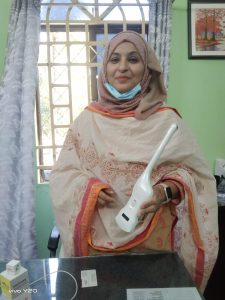 For example, a patient may present with:
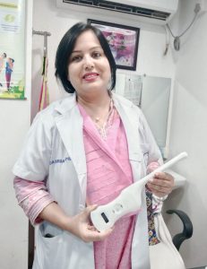

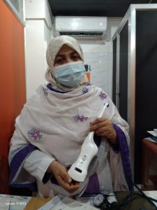
 Ultrasound is commonly used in women's healthcare to evaluate pelvic pain and pelvic masses such as ovarian cysts or uterine fibroids.
The clinical advantage of POCUS is not that it replaces every comprehensive ultrasound examination.
The advantage is that the physician can potentially obtain clinically useful information immediately.
Ultrasound is commonly used in women's healthcare to evaluate pelvic pain and pelvic masses such as ovarian cysts or uterine fibroids.
The clinical advantage of POCUS is not that it replaces every comprehensive ultrasound examination.
The advantage is that the physician can potentially obtain clinically useful information immediately.
 According to reports published by several leading Bangladeshi news and medical media outlets, physicians at the Feto-Maternal Medicine Unit of the Department of Obstetrics and Gynaecology at Dhaka Medical College Hospital successfully performed the country's first RFA procedure of this kind on 27 August 2026.
The achievement represents an important step forward for interventional medicine in Bangladesh and demonstrates the expanding clinical role of Radiofrequency Ablation technology beyond its established applications in tumor ablation, vascular intervention, pain management and other minimally invasive procedures.
According to reports published by several leading Bangladeshi news and medical media outlets, physicians at the Feto-Maternal Medicine Unit of the Department of Obstetrics and Gynaecology at Dhaka Medical College Hospital successfully performed the country's first RFA procedure of this kind on 27 August 2026.
The achievement represents an important step forward for interventional medicine in Bangladesh and demonstrates the expanding clinical role of Radiofrequency Ablation technology beyond its established applications in tumor ablation, vascular intervention, pain management and other minimally invasive procedures.
 Zhejiang CuraWay Medical Technology Co., Ltd. is a medical device manufacturer specializing in minimally invasive technologies. Its product portfolio includes Tumor Radiofrequency Ablation Systems, Endovenous Radiofrequency Ablation Systems, biopsy solutions and other interventional technologies.
CuraWay's radiofrequency technology includes dedicated RF generators and specialized electrodes designed for different clinical applications. U.S. FDA records also identify Zhejiang CuraWay Medical Technology Co., Ltd. as the applicant for its Radiofrequency Generator System and Cura RF Electrode.
Its tumor-ablation electrode portfolio includes multiple percutaneous radiofrequency ablation probes registered for commercial distribution.
Zhejiang CuraWay Medical Technology Co., Ltd. is a medical device manufacturer specializing in minimally invasive technologies. Its product portfolio includes Tumor Radiofrequency Ablation Systems, Endovenous Radiofrequency Ablation Systems, biopsy solutions and other interventional technologies.
CuraWay's radiofrequency technology includes dedicated RF generators and specialized electrodes designed for different clinical applications. U.S. FDA records also identify Zhejiang CuraWay Medical Technology Co., Ltd. as the applicant for its Radiofrequency Generator System and Cura RF Electrode.
Its tumor-ablation electrode portfolio includes multiple percutaneous radiofrequency ablation probes registered for commercial distribution.