Point-of-Care Ultrasound in Obstetrics & Gynaecology: Why TVS + Convex Ultrasound Is a Powerful Combination
Bringing Ultrasound Closer to the Patient
Ultrasound has always been central to Obstetrics and Gynaecology. What is changing today is where, when and how ultrasound is being used.
With the development of portable and handheld ultrasound technology, an OB/Gynae physician no longer has to think of ultrasound only as a separate investigation performed in a dedicated imaging room.
Point-of-Care Ultrasound (POCUS) brings real-time imaging directly to the consultation room, bedside, emergency department, labour ward or clinic.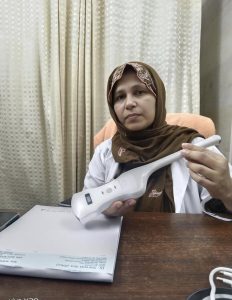
For an Obstetrician and Gynaecologist, one particularly practical configuration is a combination of:
TVS — Transvaginal Ultrasound
and
Convex — Transabdominal Ultrasound
Together, these two scanning approaches can support a wide range of focused obstetric and gynaecological assessments throughout different stages of a woman’s care.
International guidance increasingly recognizes the role of POCUS in Obstetrics and Gynaecology. In 2025, the International Society of Ultrasound in Obstetrics and Gynecology (ISUOG) published dedicated practice guidelines addressing appropriate use of POCUS in Ob/Gyn clinical scenarios, particularly when immediate bedside sonographic evaluation can assist patient management.
What Is Point-of-Care Ultrasound?
Point-of-Care Ultrasound is a focused ultrasound examination performed by the treating healthcare professional at or near the point of patient care.
Instead of sending every patient away for imaging before making the next clinical decision, a trained physician can use POCUS as an extension of the clinical examination.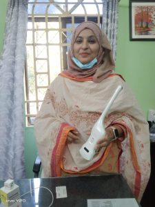
For example, a patient may present with:
- Early pregnancy with lower abdominal pain
- Vaginal bleeding
- Uncertain gestational age
- Pelvic pain
- Suspected pelvic mass
- Need to confirm fetal presentation
- Need for a rapid assessment of fetal cardiac activity
- Concern about fluid or another clinically relevant finding
A focused ultrasound examination may provide immediate additional information that helps the clinician determine the appropriate next step.
POCUS has particular value because real-time sonographic findings can immediately be interpreted alongside the patient’s symptoms, examination and clinical history.
Why OB/Gynae Needs Both TVS and Convex Ultrasound
There is no single ultrasound approach that is ideal for every Obstetric and Gynaecological patient.
This is why having both Transvaginal and Transabdominal ultrasound capability can be valuable.
The American College of Obstetricians and Gynecologists (ACOG) describes both approaches as standard ways of performing pelvic ultrasound. The appropriate approach depends on the structures that need to be visualized and the clinical reason for the examination.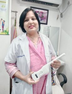
TVS — Closer Imaging for Pelvic and Early Pregnancy Assessment
A transvaginal probe can provide high-resolution imaging of structures within the pelvis because the transducer is positioned relatively close to the uterus, endometrium, cervix and adnexa.
TVS is particularly valuable when clinicians require a closer view of pelvic anatomy.
It can support assessment of areas such as:
- Uterus
- Endometrium
- Cervix
- Ovaries
- Adnexal regions
- Early intrauterine pregnancy
- Early fetal structures
- Pelvic masses
- Selected fertility-related assessments
Convex — A Broader Transabdominal View
A convex probe is placed over the abdomen and provides a broader field of view with deeper penetration.
This makes it useful across multiple phases of obstetric assessment and for broader pelvic imaging.
Depending on gestational age, clinical indication and physician training, the convex probe may support focused evaluation of:
- Pregnancy location and general overview

- Fetal presence and cardiac activity
- Fetal position or presentation
- Fetal movement
- Basic fetal measurements where appropriate
- Placental location
- Amniotic fluid assessment
- Uterine overview
- Larger pelvic structures
- Abdominal and pelvic assessment
Together, TVS + Convex creates a versatile ultrasound toolkit for the OB/Gynae physician.
1. POCUS in Early Pregnancy
Early pregnancy is one of the situations in which rapid access to ultrasound can be particularly useful.
A woman may come to an OB/Gynae clinic with:
“My pregnancy test is positive, but I have pain.”
or
“I am pregnant and experiencing bleeding.”
These presentations may require timely assessment.
A trained clinician may use focused ultrasound to obtain information relevant to questions such as: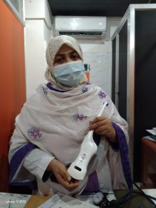
- Is an intrauterine gestational sac visualized?
- Is an embryo visualized when expected?
- Is fetal cardiac activity identified?
- Are there concerning adnexal findings?
- Is there free pelvic fluid?
- Does the clinical picture require urgent referral or comprehensive imaging?
ACOG notes that ultrasound is used during pregnancy for several purposes, including assessment of fetal health and development and evaluation when ectopic pregnancy is a concern.
In early pregnancy, the TVS probe can be especially important because of the proximity of the transducer to the pelvic organs.
2. Pelvic Pain: Seeing While Evaluating
Pelvic pain can have multiple possible causes.
History and physical examination remain fundamental, but immediate ultrasound information may help the treating physician refine the clinical picture.
A focused TVS examination can provide information about the:
Uterus → Endometrium → Ovaries → Adnexa → Pelvic cavity
It may reveal findings that require further investigation or formal imaging.
Ultrasound is commonly used in women’s healthcare to evaluate pelvic pain and pelvic masses such as ovarian cysts or uterine fibroids.
The clinical advantage of POCUS is not that it replaces every comprehensive ultrasound examination.
The advantage is that the physician can potentially obtain clinically useful information immediately.
3. Abnormal Uterine Bleeding
Abnormal uterine bleeding is another common reason women visit a Gynaecologist.
In appropriate patients, transvaginal ultrasound can assist in evaluating the uterus and endometrium as part of the overall clinical work-up.
Depending on the clinical situation, ultrasound may provide information relating to:
- Endometrial appearance
- Uterine morphology
- Fibroids
- Other structural abnormalities
- Ovarian or adnexal findings
ISUOG’s 2026 consensus guidance specifically addresses sonographic assessment of the endometrium and standardized approaches to performing and reporting gynecological ultrasound examinations.
POCUS findings should always be interpreted within the complete clinical context, with formal diagnostic imaging or additional investigation arranged when necessary.
4. Fibroids and Other Uterine Findings
Uterine fibroids are frequently encountered in Gynaecology.
Ultrasound can help physicians assess the uterus and identify findings compatible with fibroids.
TVS may provide close pelvic visualization, while a convex abdominal probe can offer a broader view—particularly when the uterus is enlarged or a wider field is required.
This demonstrates one of the key advantages of having both TVS and Convex capability in the same portable ultrasound platform.
The clinician can choose the scanning approach according to the patient and the clinical question.
5. Ovarian and Adnexal Assessment
The ovaries and adnexa represent another major area of gynaecological ultrasound.
TVS can help visualize ovarian morphology and identify adnexal abnormalities requiring further evaluation.
Ultrasound may be used to evaluate:
- Ovarian cysts
- Adnexal masses
- Pelvic pain
- Selected infertility-related questions
- Other pelvic abnormalities
ACOG identifies pelvic masses, infertility evaluation and monitoring of infertility treatment among the established uses of ultrasound in women’s healthcare.
Complex ovarian or adnexal lesions, however, may require a comprehensive specialist ultrasound examination, Doppler assessment and/or further imaging rather than relying on a limited POCUS examination alone.
6. Fetal Cardiac Activity at the Point of Care
One of the most clinically meaningful moments in obstetric practice is confirming fetal cardiac activity.
Depending on gestational age, a physician may use TVS or transabdominal imaging to evaluate fetal cardiac activity.
For patients presenting with pain, bleeding or pregnancy-related anxiety, being able to perform an appropriate focused scan during the clinical encounter may provide valuable immediate information.
It can also help clinicians decide whether further formal evaluation is required.
7. Fetal Presentation
As pregnancy progresses, a Convex probe becomes particularly useful.
A focused transabdominal scan can help an appropriately trained OB/Gynae physician determine fetal presentation, such as:
Cephalic
Breech
or
Transverse / other lie
This may be valuable during antenatal visits, late-pregnancy assessment or labour-related evaluation.
For many clinical questions, having ultrasound immediately available can make it easier to combine physical examination findings with real-time imaging.
8. Placental Location
Placental location is an important part of obstetric ultrasound assessment.
A Convex probe can provide a broad transabdominal view of the uterus and placenta.
In certain situations, additional transvaginal assessment may be necessary to evaluate the relationship between the placenta and cervix more accurately.
This again illustrates why TVS and Convex should not always be considered competing probes.
They complement one another.
The appropriate approach depends on gestational age, anatomy, clinical question and operator expertise.
9. Amniotic Fluid Assessment
Transabdominal ultrasound may also assist trained clinicians in focused evaluation of amniotic fluid.
When clinically indicated, ultrasound findings can contribute to the assessment of whether fluid volume appears appropriate or whether a formal obstetric ultrasound evaluation is required.
Current ISUOG guidance for later-pregnancy ultrasound includes evaluation of fetal presentation, placental location, fetal biometry and amniotic fluid as components of obstetric sonographic assessment.
10. A Powerful Tool for Emergency OB/Gynae Assessment
Consider an emergency scenario:
A pregnant woman arrives with abdominal pain and vaginal bleeding.
Traditionally, the physician might perform the examination and then wait for access to an ultrasound department.
With a handheld POCUS device available at the bedside, a trained clinician may be able to obtain focused sonographic information immediately and combine it with vital signs, history and examination.
This does not mean that every handheld ultrasound examination provides a final diagnosis.
But it can help answer an important question:
“What do I need to know right now to decide the next clinical step?”
This question-oriented nature is one of the central strengths of Point-of-Care Ultrasound.
TVS + Convex: Two Perspectives, One Clinical Workflow
Think of the two probes as providing complementary perspectives.
TVS
Closer. Higher-resolution pelvic assessment.
Particularly useful for:
- Early pregnancy
- Uterus
- Endometrium
- Cervix
- Ovaries
- Adnexa
- Pelvic pathology
Convex
Wider. Deeper transabdominal assessment.
Particularly useful for:
- Obstetric overview
- Later pregnancy
- Fetal position
- Placental assessment
- Amniotic fluid
- Broader uterine and pelvic evaluation
TVS + Convex = A More Complete Point-of-Care Solution for OB/Gynae
Rather than carrying separate ultrasound systems, a dual-probe handheld solution can allow clinicians to choose the appropriate scanning approach based on the patient in front of them.
Why Handheld Wireless Ultrasound?
The transformation is not simply about making an ultrasound machine smaller.
It is about changing access to imaging.
A handheld wireless ultrasound can potentially fit into the everyday clinical workflow:
Patient arrives → Clinical history → Physical examination → Focused POCUS → Clinical decision
Instead of:
Patient arrives → Examination → Imaging request → Ultrasound department → Waiting → Report → Return to physician
Not every patient or clinical question can follow the first pathway, and comprehensive diagnostic examinations remain essential when indicated.
But for appropriately selected focused examinations, portability can make ultrasound much more accessible at the point of care.
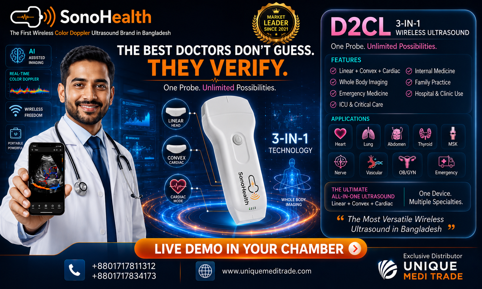
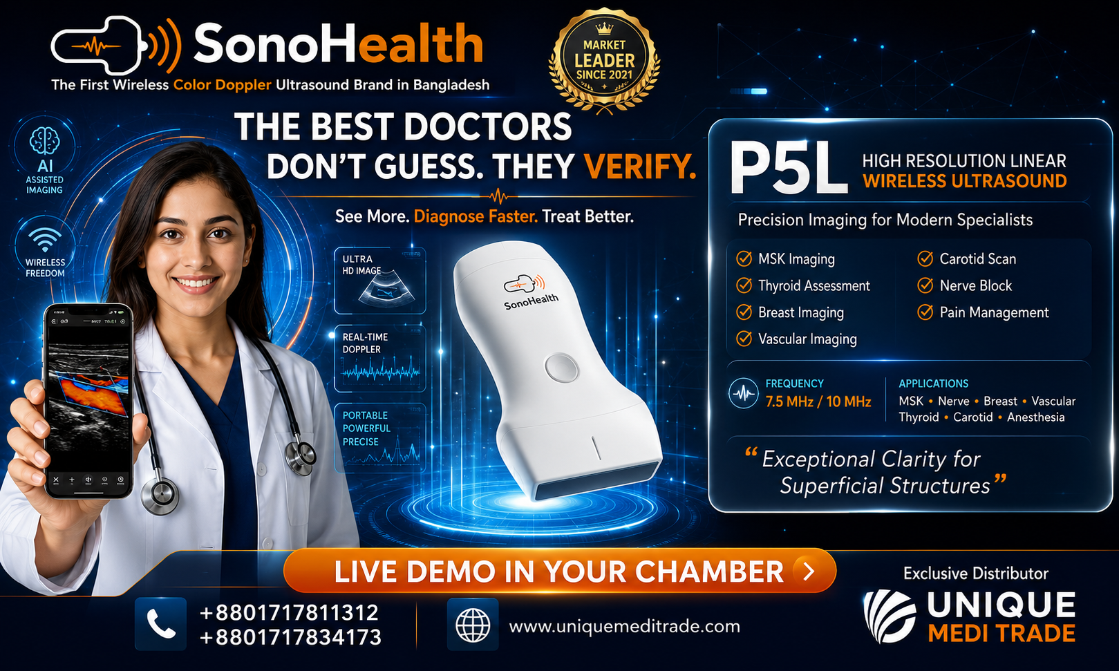
SonoHealth Handheld Wireless Ultrasound — TVS + Convex
For OB/Gynae specialists looking to integrate point-of-care ultrasound into everyday clinical practice, SonoHealth Handheld Wireless Ultrasound with TVS + Convex configuration provides two important ultrasound approaches within a highly portable platform.
TVS Probe
Designed for close-range transvaginal imaging for applications such as early pregnancy and pelvic assessment.
Convex Probe
Designed for transabdominal imaging, providing greater depth and a broader field of view for obstetric and pelvic applications.
The handheld format allows the ultrasound system to work with compatible smart devices, making imaging more accessible in environments such as:
- Private consultation chambers
- OB/Gynae clinics
- Hospitals
- Labour wards
- Emergency departments
- Bedside examinations
- Rural and peripheral healthcare settings
- Mobile clinical services
The objective is simple:
Bring ultrasound to the physician instead of always bringing the patient to the ultrasound machine.
From “I Think” to “I Can See”
Clinical medicine will always depend on history, examination, experience and professional judgment.
POCUS adds something powerful to that process:
Real-time visualization.
When appropriately trained physicians can see relevant anatomy at the point of care, they may be able to integrate that information immediately with the clinical picture.
That is why handheld ultrasound should not simply be viewed as a smaller ultrasound machine.
For many physicians, it can become an extension of the clinical examination.
POCUS Does Not Replace Comprehensive Diagnostic Ultrasound
This distinction is important.
Point-of-Care Ultrasound and comprehensive diagnostic ultrasound are not necessarily the same examination.
POCUS is generally performed to answer a focused clinical question relevant to immediate patient management.
A comprehensive diagnostic ultrasound may require:
- Detailed standardized imaging protocols
- Multiple measurements
- Advanced Doppler assessment
- Detailed fetal anatomical evaluation
- Specialist interpretation
- Structured documentation
- Follow-up examination
AIUM practice parameters emphasize defined minimum criteria, appropriate examination techniques and documentation for high-quality diagnostic ultrasound.
Therefore, suspicious, complex or inconclusive findings identified during POCUS should be referred for appropriate comprehensive assessment.
The goal of POCUS is not to replace specialists. It is to give appropriately trained clinicians another tool for timely clinical decision-making.
Training Matters
Buying an ultrasound device does not automatically create a POCUS program.
Clinical value comes from the combination of:
Technology + Physician Training + Scanning Technique + Clinical Knowledge + Appropriate Patient Selection
OB/Gynae physicians using handheld ultrasound should receive appropriate training in image acquisition, anatomy, interpretation, documentation, infection prevention and the limitations of focused ultrasound.
ISUOG’s dedicated Ob/Gyn POCUS guideline reflects the increasing importance of establishing appropriate standards as point-of-care ultrasound becomes more widely adopted.
The Future of OB/Gynae Ultrasound Is Becoming More Accessible
Ultrasound technology has evolved from large machines confined to dedicated rooms to compact systems that can be carried to the bedside.
For Obstetricians and Gynaecologists, this evolution has special significance because ultrasound is already deeply integrated into the specialty.
A TVS + Convex handheld wireless ultrasound gives the clinician two complementary imaging approaches:
TVS for closer pelvic visualization.
Convex for broader transabdominal and obstetric visualization.
Together, they can create a practical point-of-care imaging solution for modern OB/Gynae practice.
See the Patient. Scan at the Point of Care. Decide with More Information.
The future of women’s healthcare is not simply about more technology.
It is about putting the right technology in the right hands at the right moment.
Point-of-Care Ultrasound can help bring real-time imaging closer to the patient—and closer to the physician making the clinical decision.
SonoHealth Handheld Wireless Ultrasound
TVS + Convex
Portable Imaging for Modern OB/Gynae Practice
See More. Diagnose Faster. Treat Better.
For product information, physician demonstration, clinical discussion or quotation:
Unique Medi Trade
Handheld Wireless Ultrasound Solutions in Bangladesh
Contact: +880 1717-811312
Medical Disclaimer
This article is intended for professional and educational information only. POCUS should be performed by appropriately trained healthcare professionals and used according to applicable clinical guidelines, institutional protocols and local regulations. A focused POCUS examination should not replace comprehensive diagnostic ultrasound or specialist evaluation when these are clinically indicated.