Point-of-Care Ultrasound (POCUS) in Vascular Medicine and Vascular Surgery: See the Vessel, Understand the Flow, Guide the Procedure
Bringing Vascular Ultrasound Directly to the Point of Care
Vascular medicine is a specialty built around anatomy, circulation and blood flow.
A patient may present with:
Leg swelling.
Sudden limb pain.
Varicose veins.
Suspected Deep Vein Thrombosis (DVT).
Poor peripheral circulation.
A difficult vascular access.
An arteriovenous fistula problem.
Or a vascular surgeon may need to perform an ultrasound-guided endovenous intervention.
In all of these situations, seeing the vessel can provide valuable information that physical examination alone cannot provide.
This is where Point-of-Care Ultrasound (POCUS) is becoming increasingly relevant.
POCUS allows an appropriately trained physician to perform a focused ultrasound examination at the bedside, outpatient chamber, procedure room or operating environment and immediately integrate the findings into clinical decision-making.
A recent review of vascular POCUS describes its expanding use beyond procedural guidance, particularly for evaluation of lower-extremity deep venous thrombosis and the abdominal aorta.
For vascular specialists, however, its potential goes even further.
Vein. Artery. Flow. Needle. Catheter.
POCUS puts vascular anatomy directly into the physician’s hands.
What Is Vascular POCUS?
Point-of-Care Ultrasound is generally a focused examination performed and interpreted by the treating clinician to answer a specific clinical question.
For a vascular specialist, those questions may include:
- Is this vein compressible?
- Is proximal DVT suspected?
- Where is the target vein?
- Where is the artery?
- What is the diameter and depth of the vessel?
- Which vessel is most appropriate for access?
- Is blood flow visible with Color Doppler?
- Is there a superficial venous abnormality?
- Is the abdominal aorta enlarged?
- Is an AV fistula accessible for cannulation?
- Is there a collection, hematoma or suspected pseudoaneurysm?
- Where should an endovenous catheter or needle be positioned?
- What structures should be avoided during an intervention?
This question-focused approach is different from a complete vascular laboratory duplex examination.
POCUS is designed primarily to provide immediate, targeted information.
Why POCUS Is Particularly Relevant to Vascular Specialists
The vascular specialist works with structures that are highly suitable for ultrasound.
Many important vessels are relatively superficial and can be clearly differentiated using B-mode and Doppler imaging.
With ultrasound, the physician can potentially evaluate: 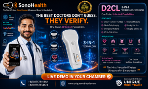
Vessel Anatomy
Location, depth, diameter and relationship with surrounding structures.
Compressibility
Particularly relevant in venous thrombosis assessment.
Blood Flow
Using Color and spectral Doppler when appropriate.
Needle Position
During vascular access and other interventions.
Surrounding Anatomy
Including nerves, muscles, soft tissue and adjacent arteries or veins.
This creates an important transition in vascular practice:
From feeling the pulse to seeing the artery.
From locating the vein by landmarks to visualizing it.
From estimating needle position to guiding it in real time.
1. Point-of-Care Ultrasound for Deep Vein Thrombosis
Deep Vein Thrombosis is one of the most established diagnostic applications of vascular POCUS.
Patients may present with:
- Unilateral leg swelling
- Leg pain
- Tenderness
- Increased limb circumference
- Risk factors for venous thromboembolism
Clinical examination alone cannot reliably confirm or exclude DVT.
Focused compression ultrasound can allow a trained physician to assess whether key proximal veins compress normally.
The fundamental concept is simple:
Normal vein → Usually compresses under appropriate probe pressure.
Thrombosed vein → May fail to compress normally.
A 2024 systematic review and meta-analysis found strong diagnostic performance for several clinician-performed POCUS protocols for DVT, although performance depends on technique, training and the specific protocol used.
A dedicated review also concluded that POCUS can have excellent diagnostic accuracy for acute proximal lower-extremity DVT when performed by well-trained users, while emphasizing the need for appropriate education and standardized training.
For a vascular physician evaluating a patient with suspected DVT, this means bedside ultrasound may provide clinically important information immediately.
2. Compression Ultrasound: A Practical Vascular POCUS Skill
Compression ultrasound is one of the fundamental skills of venous POCUS.
The physician identifies the artery and vein in transverse view and applies controlled pressure with the probe.
The examination may assess selected venous segments according to the protocol being used.
A vascular POCUS examination may incorporate:
B-Mode
for anatomy and compressibility,
Color Doppler
for visualization of flow,
and when appropriate,
Spectral Doppler
for additional hemodynamic information.
Professional ultrasound guidance for peripheral venous examinations emphasizes structured assessment and documentation for complete diagnostic studies.
This highlights an important distinction:
Focused DVT POCUS and comprehensive venous duplex ultrasound are related—but they are not always interchangeable.
If findings are uncertain, symptoms suggest distal or complex disease, or detailed venous mapping is required, a complete vascular duplex examination may still be necessary.
3. Varicose Veins and Chronic Venous Disease
Chronic venous disease is a major part of vascular practice.
Patients may present with:
- Varicose veins

- Leg heaviness
- Pain
- Swelling
- Skin pigmentation
- Venous eczema
- Lipodermatosclerosis
- Venous ulceration
Ultrasound plays a fundamental role in evaluating venous anatomy and reflux.
Current Society for Vascular Surgery, American Venous Forum and American Vein and Lymphatic Society guidelines include duplex ultrasound scanning as a central component in evaluating patients with lower-extremity varicose veins and superficial truncal reflux.
For the vascular specialist, a handheld ultrasound can be useful during the initial clinical encounter for focused questions.
For example:
Which superficial vein is visible beneath the symptomatic area?
Is the vessel patent?
What is its approximate diameter and depth?
Is obvious reflux or abnormal flow suspected?
However, treatment planning for chronic venous insufficiency generally requires a structured duplex study, including detailed reflux assessment and mapping.
POCUS can complement that workflow rather than replace it.
4. Ultrasound-Guided Endovenous Radiofrequency Ablation
One of the strongest examples of ultrasound becoming an integral part of vascular intervention is Endovenous Radiofrequency Ablation (RFA).
Endovenous thermal ablation is used in appropriately selected patients with superficial venous reflux and varicose vein disease.
During a typical ultrasound-guided procedure, ultrasound may assist the physician in:
Identifying the Target Vein 
Selecting an Access Site
Guiding Needle Entry
Guiding Guidewire and Catheter Position
Evaluating the Relationship to Deep Veins
Guiding Tumescent Anaesthesia
Monitoring the Procedure
Assessing the Treated Vein After Intervention
International consensus describes endovenous thermal ablation—including radiofrequency—as a catheter-directed, ultrasound-guided treatment for varicose vein disease.
A multidisciplinary international position statement also recommends concurrent ultrasound imaging for superficial endovenous procedures including radiofrequency ablation, endovenous laser ablation, cyanoacrylate closure and ultrasound-guided sclerotherapy.
This makes ultrasound much more than a diagnostic tool for vascular surgeons.
It becomes a procedural navigation tool.
5. Ultrasound-Guided Vascular Access
One of the most practical uses of POCUS is vascular access.
Every clinician knows the challenge:
A vein may be palpable in one patient but almost impossible to identify in another.
Ultrasound allows the operator to visualize:
Vein
Artery
Depth
Diameter
Surrounding structures
Needle
before and during cannulation.
POCUS can be used for:
- Peripheral venous access
- Central venous access
- Selected arterial access
- Difficult vascular access
- Dialysis access-related procedures
International evidence-based recommendations support ultrasound guidance for central venous cannulation and note its value in selected peripheral venous and arterial access procedures.
A review of Cochrane evidence found ultrasound-guided vascular access associated with improvements in outcomes such as first-attempt success and overall success in several studied settings, while also reducing some failed attempts and complications.
6. Seeing the Needle During Vascular Access
A major advantage of ultrasound guidance is the ability to observe needle advancement.
Two commonly used techniques are:
In-Plane
The needle advances along the ultrasound imaging plane, potentially allowing visualization of much of the needle shaft and tip.
Out-of-Plane
The needle crosses the ultrasound beam and appears as a bright echogenic point when intersecting the imaging plane.
The important principle is:
Always understand where the needle tip is.
Seeing part of the needle does not necessarily mean the operator has identified the tip.
Training in hand-eye coordination, probe positioning and needle visualization is therefore essential.
7. Arterial POCUS
Arterial ultrasound can provide valuable focused information in vascular practice.
Depending on the clinical situation and device capability, a vascular physician may use ultrasound to evaluate:
- Arterial location
- Vessel diameter
- Patency
- Pulsatility
- Calcification
- Selected flow characteristics
- Suitable arterial access sites
- Surrounding anatomy
Color Doppler can help differentiate arterial and venous flow, while spectral Doppler may provide additional hemodynamic information.
For comprehensive assessment of Peripheral Arterial Disease (PAD), however, formal vascular testing may involve:
- Ankle-Brachial Index
- Segmental pressures
- Pulse-volume recordings
- Complete arterial duplex
- CT angiography
- MR angiography
- Conventional angiography
depending on the clinical situation.
Therefore:
POCUS can answer focused arterial questions, but it should not automatically be considered a substitute for comprehensive vascular investigation.
8. Abdominal Aortic Aneurysm
The abdominal aorta is another important target for POCUS.
A focused transabdominal examination can help determine whether the aorta appears enlarged in a patient where an abdominal aortic aneurysm is suspected.
This may be particularly relevant for patients presenting with combinations such as:
Abdominal pain
Back pain
Hypotension
Syncope
and appropriate vascular risk factors.
Recent evidence continues to support the ability of trained clinicians to detect AAA with POCUS. A 2026 review of systematic reviews reported very high ranges of sensitivity and specificity for clinician-performed POCUS in diagnosing AAA.
A 2024 systematic review and meta-analysis similarly reported high diagnostic performance in emergency settings.
For a vascular specialist, however, identifying an aneurysm is only the beginning.
Detailed treatment planning may require comprehensive ultrasound, CT angiography or other vascular imaging to assess anatomy, branch vessels, morphology and suitability for intervention.
9. Pseudoaneurysm Assessment
Pseudoaneurysm can occur following arterial puncture, catheterization, trauma or vascular procedures.
Ultrasound can help evaluate a suspicious groin or access-site swelling and identify vascular flow patterns suggesting a pseudoaneurysm.
Depending on the case and operator expertise, ultrasound may also support selected image-guided treatment.
EFSUMB guidance on ultrasound-guided vascular interventions includes ultrasound for detection of vascular-access complications and ultrasound-guided treatment of arterial pseudoaneurysms.
This is another example of POCUS moving beyond simple diagnosis into intervention.
10. Arteriovenous Fistula and Dialysis Access
Patients receiving haemodialysis depend on reliable vascular access.
POCUS can be used for focused evaluation of an AV fistula or graft.
The physician may assess questions such as:
- Where is the fistula?
- How deep is the vessel?
- What is its diameter?
- Is there visible flow?
- Is there an obvious abnormality?
- Where is the most appropriate area for cannulation?
A review of vascular-access POCUS emphasizes that its goal is often to answer focused bedside questions rather than reproduce a complete fistula or graft duplex examination.
A broader review found POCUS has been used for AV fistula maturation assessment, identification of abnormalities, difficult cannulation and selection of alternative cannulation sites.
This distinction perfectly illustrates the philosophy of POCUS:
Ask a focused question.
Scan.
Obtain immediate information.
Decide whether additional investigation is required.
11. Color Doppler: Seeing More Than Anatomy
B-mode ultrasound shows anatomy.
Color Doppler adds information about blood flow.
For vascular physicians, this can be particularly valuable.
Color Doppler may help identify:
- Vessel flow
- Flow direction
- Areas of abnormal flow
- Relationship between vessels
- Possible vascular abnormalities
- Patency during focused assessment
Spectral Doppler can further provide information about flow velocity and waveform morphology when the equipment and operator training support it.
For advanced vascular assessment, Doppler capability becomes especially important because vascular medicine is not simply about seeing vessels.
It is about understanding circulation.
12. POCUS Before a Vascular Procedure
Imagine preparing for an intervention.
Before puncturing the skin, the physician can scan the area and ask:
Where exactly is the artery?
Where is the vein?
How deep is the vessel?
What is its diameter?
Is there another vessel nearby?
Is there calcification?
Is this the best access point?
What is the safest needle trajectory?
This brief pre-procedural scan can transform a landmark-based procedure into an image-informed procedure.
International vascular-access recommendations support ultrasound screening before cannulation and real-time needle guidance in appropriate settings.
13. POCUS During the Procedure
The value continues after skin puncture.
Real-time ultrasound can potentially allow the operator to see:
Needle → Vessel → Guidewire → Catheter
depending on the procedure.
For endovenous treatment, ultrasound may also be used to observe surrounding anatomy and assist with proper treatment-zone positioning.
This creates a major difference between:
“I believe the device is in the correct location.”
and
“I can visualize where it is.”
14. POCUS After the Procedure
Focused ultrasound may also contribute to immediate post-procedure assessment.
Depending on the intervention, the physician may look for:
- Vessel patency
- Treatment response
- Hematoma
- Suspected access-site complication
- Pseudoaneurysm
- Local fluid collection
- Other relevant findings
More comprehensive post-treatment surveillance may still require a formal vascular duplex study according to the procedure and clinical guideline.
Duplex ultrasound remains a fundamental part of assessment following many venous interventions.
POCUS and Endovenous RFA: A Natural Combination
For a vascular practice performing Radiofrequency Ablation for Varicose Veins, ultrasound and RFA naturally work together.
A simplified clinical workflow may look like:
Clinical Assessment
↓
Duplex Evaluation
↓
Treatment Planning
↓
Ultrasound-Guided Venous Access
↓
Catheter Positioning
↓
Tumescent Anaesthesia Under Ultrasound Guidance
↓
Radiofrequency Ablation
↓
Ultrasound Assessment
Ultrasound provides visualization.
Radiofrequency provides the therapeutic energy.
Together, these technologies support modern minimally invasive treatment of appropriately selected superficial venous disease.
Which Ultrasound Probe Does a Vascular Specialist Need?
Linear Probe — The Essential Vascular Probe
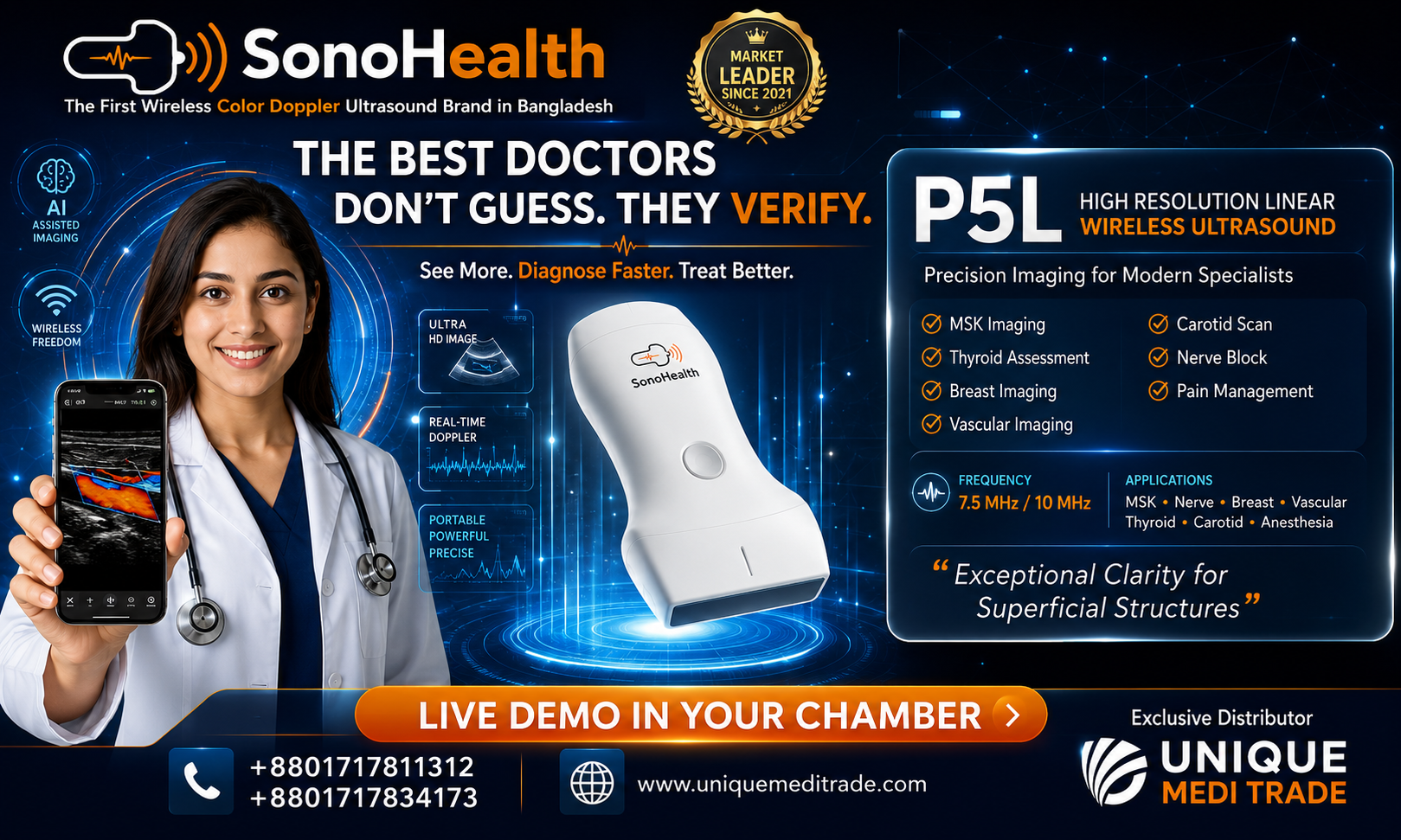
For most superficial vascular applications, a high-frequency Linear probe is particularly important.
It is useful for:
- DVT assessment
- Superficial veins
- Vascular access
- Carotid and other superficial vessels
- Varicose veins
- Endovenous procedures
- AV fistula
- Peripheral arteries
- Needle guidance
- Soft-tissue assessment
The rectangular image and high-frequency capability make the linear transducer particularly suitable for superficial anatomy.
Why a Convex Probe Can Also Be Useful
Not every vessel is superficial.
For deeper structures, the Convex probe can provide greater penetration and a wider field of view.
Potential applications include:
- Abdominal aorta
- Iliac-region assessment
- Deep abdominal structures
- Selected deep vascular anatomy
- Abdominal POCUS
This means that a vascular specialist may benefit from having:
Linear for superficial vascular imaging
and
Convex for deeper abdominal vascular assessment.
Why Handheld Wireless Ultrasound Makes Sense in Vascular Practice
Traditional vascular ultrasound systems remain essential for detailed duplex examinations.
But a vascular surgeon does not perform every clinical assessment inside a vascular laboratory.
The specialist may be working in:
- Consultation chamber
- Vascular clinic
- Hospital ward
- Emergency department
- Procedure room
- Operating theatre
- Dialysis unit
- Bedside
- Peripheral healthcare facility
A handheld wireless ultrasound can travel with the physician.
It can potentially connect to a compatible smartphone or tablet and provide immediate imaging when a focused clinical question arises.
The workflow becomes:
Examine → Scan → Visualize → Decide → Intervene when appropriate.
This is the philosophy behind Point-of-Care Ultrasound.
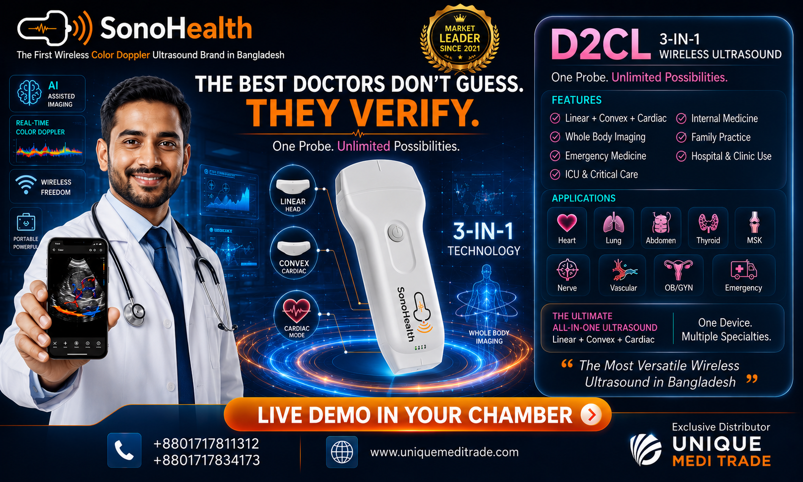
SonoHealth Handheld Wireless Ultrasound for Vascular POCUS
For vascular specialists looking to introduce portable ultrasound into everyday practice, SonoHealth Handheld Wireless Ultrasound provides a practical point-of-care imaging platform.
Depending on the configuration, it can support:
Vascular Imaging
Focused examination of veins and arteries.
Color Doppler
Visualization of vascular flow.
DVT Assessment
Focused compression ultrasound in appropriately selected patients.
Vascular Access
Real-time ultrasound guidance for needle placement.
Endovenous Procedures
Support for image-guided vascular intervention.
AV Fistula Assessment
Focused evaluation and access guidance.
Abdominal Vascular Assessment
With an appropriate deeper-penetration transducer.
Needle Guidance
For selected vascular procedures.
For many vascular applications, the combination of a high-frequency Linear probe + Color Doppler + needle-guidance capability can be particularly useful.
Adding a Convex probe expands the potential range toward deeper abdominal and aortic imaging.
POCUS Does Not Replace a Complete Vascular Duplex Study
This is one of the most important principles when adopting handheld ultrasound.
A focused POCUS examination and a comprehensive vascular duplex examination are not necessarily the same thing.
A complete vascular laboratory examination may require:
- Detailed vessel mapping
- Standardized Doppler waveforms
- Velocity measurements
- Reflux testing
- Spectral Doppler analysis
- Multiple standardized anatomical segments
- Formal interpretation
- Structured reporting
- Quality-control protocols
Professional vascular guidelines continue to rely heavily on comprehensive duplex ultrasound for conditions such as chronic venous disease and treatment planning.
Therefore, POCUS should be considered:
A focused clinical tool.
A procedural guidance tool.
An extension of vascular examination.
not automatically a replacement for comprehensive vascular imaging.
Training Matters
Ultrasound is operator-dependent.
A sophisticated ultrasound device is only useful when the physician understands what is being displayed.
Vascular POCUS training should include:
- Ultrasound physics
- Probe orientation
- Vascular anatomy
- Artery-versus-vein identification
- Compression technique
- B-mode optimization
- Color Doppler
- Spectral Doppler where applicable
- Needle visualization
- In-plane and out-of-plane approaches
- Recognition of artifacts
- Recognition of DVT findings
- Recognition of vascular-access complications
- Understanding when comprehensive imaging is required
Published evidence on DVT POCUS repeatedly emphasizes that good diagnostic performance depends on adequately trained operators.
Technology + Training + Clinical Knowledge = Effective Vascular POCUS.
The Future of Vascular Medicine Is Increasingly Visual
Vascular specialists have always relied heavily on clinical examination.
They inspect the limb.
They palpate pulses.
They evaluate edema.
They assess skin changes.
They listen for bruits.
POCUS adds another dimension:
They can see the vessel.
They can see flow.
They can see the needle.
They can see the target before intervention.
This does not replace clinical examination.
It strengthens it.
From Palpation to Visualization
A handheld ultrasound can change the everyday vascular workflow.
Instead of:
“I think the vein is here.”
the physician may be able to say:
“I can see the vein.”
Instead of:
“The vessel should be approximately this deep.”
“I can measure its depth.”
Instead of:
“The needle should be approaching the vessel.”
“I can follow the needle toward the target.”
Instead of relying solely on external anatomy:
The vascular specialist can visualize what lies underneath.
That is the practical value of POCUS.
See the Vessel. Understand the Flow. Guide the Procedure.
Point-of-Care Ultrasound is helping bring real-time vascular imaging closer to the physician and closer to the patient.
For vascular specialists, its potential spans:
DVT Assessment
Venous Examination
Arterial Assessment
Vascular Access
Varicose Vein Procedures
Endovenous RFA
AV Fistula Assessment
Aortic POCUS
Needle Guidance
and selected post-procedural assessments.
The future is not simply about smaller ultrasound machines.
It is about having relevant imaging available when the clinical decision is being made.
SonoHealth Handheld Wireless Ultrasound
Point-of-Care Ultrasound for Modern Vascular Practice
Vascular Imaging • Color Doppler • DVT Assessment • Vascular Access • Endovenous Procedures • Needle Guidance
See the Vessel. Understand the Flow. Guide with Precision.
For vascular surgeons, vascular specialists, hospitals and vascular centers interested in integrating handheld Point-of-Care Ultrasound into clinical practice:
Unique Medi Trade
POCUS Solutions • Live Demonstration • Clinical Training Support • Technical Support • Endovenous RFA Solutions
Contact: +880 1717-811312
Unique Medi Trade — Advancing Image-Guided Vascular Care in Bangladesh
Medical Disclaimer
This article is intended for healthcare-professional education and general information. Point-of-Care Ultrasound should be performed by appropriately trained healthcare professionals within their scope of practice and according to relevant clinical guidelines and institutional protocols.
Focused POCUS should not replace a comprehensive vascular duplex examination, CT angiography, MR angiography, conventional angiography or specialist vascular imaging when these are clinically indicated. Clinical decisions should be based on the complete patient assessment rather than an isolated POCUS finding.
SEO Information
SEO Title:
Point-of-Care Ultrasound (POCUS) in Vascular Medicine & Vascular Surgery
Alternative SEO Title:
POCUS for Vascular Surgeons: DVT, Vascular Access, Varicose Veins & Endovenous Procedures
Meta Description:
Discover how Point-of-Care Ultrasound (POCUS) supports vascular specialists in DVT assessment, vascular access, venous and arterial imaging, endovenous RFA, AV fistula assessment and image-guided procedures.
Suggested URL Slug:
/point-of-care-ultrasound-pocus-in-vascular-surgery/
Target Keywords:
Point-of-Care Ultrasound Vascular, POCUS Vascular Surgery, vascular ultrasound, handheld vascular ultrasound, DVT ultrasound, ultrasound-guided vascular access, varicose vein ultrasound, Endovenous RFA, Color Doppler ultrasound, vascular surgeon ultrasound, SonoHealth Handheld Ultrasound, POCUS Bangladesh
Suggested Featured Image Text:
POINT-OF-CARE ULTRASOUND IN VASCULAR MEDICINE
See the Vessel. Understand the Flow. Guide with Precision.
SonoHealth Handheld Wireless Ultrasound