Point-of-Care Ultrasound (POCUS): Bringing Real-Time Imaging to the Bedside
Introduction
Modern medicine increasingly depends on rapid, accurate, and clinically relevant information. Traditionally, physicians have relied on history taking, physical examination, laboratory testing, and formal imaging such as radiology ultrasound, CT, MRI, and echocardiography to reach a diagnosis. While these remain essential, many clinical decisions must be made immediately at the bedside.
This is where
Point-of-Care Ultrasound, commonly known as POCUS, has become increasingly important.
POCUS refers to the use of ultrasound by the treating clinician at or near the patient's bedside to answer specific clinical questions, guide procedures, and support real-time decision-making. Instead of sending every patient to an imaging department, clinicians trained in POCUS can perform focused ultrasound examinations during the clinical encounter.
POCUS is now used in emergency medicine, internal medicine, critical care, anesthesia, pain management, cardiology, surgery, orthopedics, obstetrics and gynecology, urology, primary care, and several other specialties.
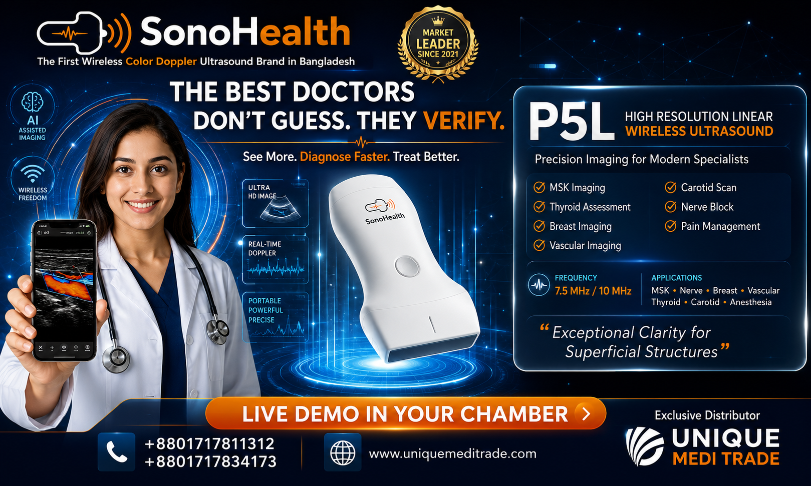
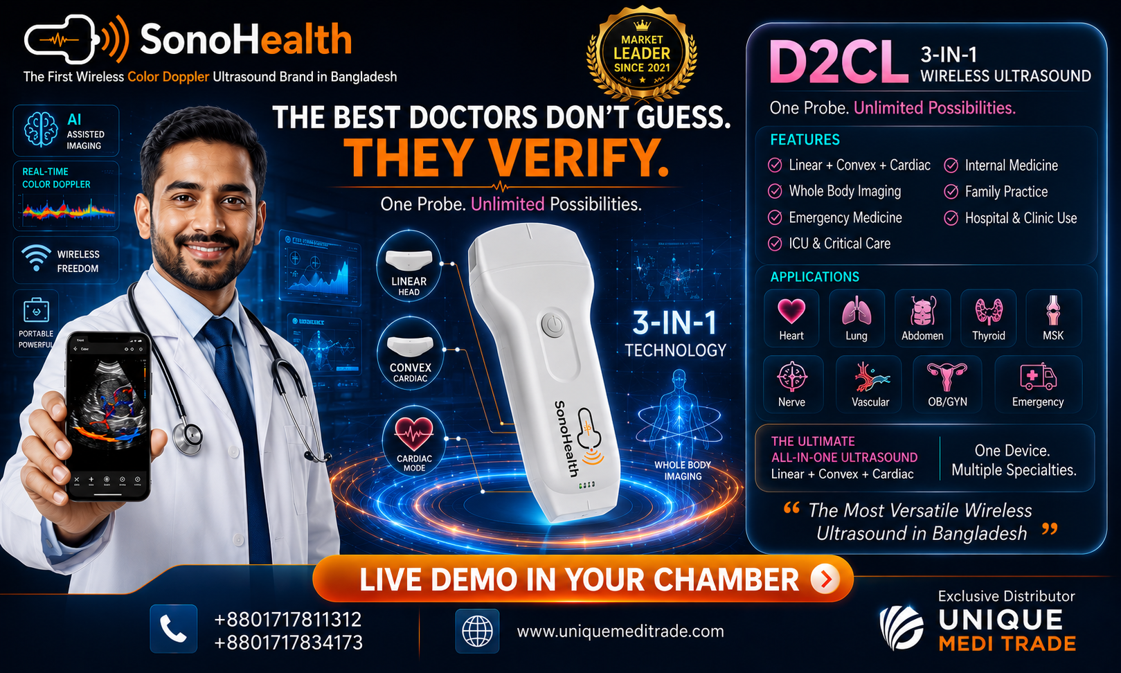
Its growing popularity has been accelerated by improvements in portable and handheld ultrasound technology. Modern devices can connect with smartphones or tablets, making diagnostic imaging increasingly accessible at the point of care.
POCUS is not simply a smaller version of traditional ultrasound. It represents a different clinical approach:
focused imaging performed to answer focused questions in real time.
What Is Point-of-Care Ultrasound?
Point-of-Care Ultrasound is a focused ultrasound examination performed and interpreted by the clinician directly responsible for the patient's care.
The word "point-of-care" is important. The scan is usually performed where the patient is being evaluated or treated, such as:
- Emergency departments
- Intensive care units
- Hospital wards
- Outpatient clinics
- Operating rooms
- Procedure rooms
- Ambulances
- Rural health centers
- Remote medical facilities
- Home-care environments
The purpose of POCUS is generally not to perform a complete radiological examination of an entire organ system. Instead, the clinician uses ultrasound to answer a specific question.
For example:
Does this patient have a pleural effusion?
Is there evidence of pulmonary edema?
Is there pericardial fluid?
Is the urinary bladder significantly distended?
Is there free fluid in the abdomen?
Where is the target nerve for an ultrasound-guided injection?
Where is the safest site for vascular access?
These focused questions can significantly influence immediate clinical management.
POCUS and the Modern Physical Examination
For generations, physicians have used four fundamental techniques during physical examination:
Inspection, palpation, percussion, and auscultation.
POCUS is increasingly considered an extension of these traditional examination skills.
A physician may suspect fluid overload after listening to the lungs and examining peripheral edema. Lung ultrasound can provide additional real-time information by demonstrating ultrasound patterns associated with interstitial fluid.
Similarly, a physician may suspect ascites clinically and then quickly confirm the presence and approximate distribution of fluid using bedside ultrasound.
This does not mean that ultrasound replaces clinical examination. Instead, POCUS can strengthen clinical assessment by adding visual information.
The concept can be summarized simply:
Look. Listen. Examine. Scan. Decide.
How POCUS Differs from Conventional Ultrasound
POCUS and formal diagnostic ultrasound are related but not identical.
A traditional ultrasound examination is usually comprehensive. It may be performed by a sonographer, radiologist, cardiologist, or another imaging specialist following an established imaging protocol. A formal report is then produced.
POCUS is usually focused.
The clinician may perform only the views required to answer a specific clinical question.
For example, a formal echocardiogram may include detailed assessment of cardiac chambers, valves, Doppler measurements, ventricular function, pressure estimates, and multiple standardized views.
A focused cardiac POCUS examination may instead seek to determine whether there is:
- Severe global ventricular dysfunction
- Significant pericardial effusion
- Right ventricular enlargement
- Marked volume depletion or overload
- Major abnormality requiring urgent evaluation
Therefore, POCUS should be considered
complementary to comprehensive diagnostic imaging rather than a replacement for it.
When the clinical question requires detailed imaging, formal ultrasound, echocardiography, CT, MRI, or specialist evaluation remains necessary.
Why POCUS Is Becoming Important
One of the strongest advantages of POCUS is speed.
A patient with acute shortness of breath may have several possible causes, including pneumonia, pulmonary edema, pleural effusion, pneumothorax, cardiac dysfunction, or other conditions.
Traditionally, the clinician may need to wait for chest radiography, formal ultrasound, laboratory results, or additional imaging.
With appropriate training, POCUS can provide useful bedside information within minutes.
This can be particularly valuable when patients are unstable.
Other important advantages include:
Immediate Clinical Information
POCUS provides information while the physician is actively evaluating the patient.
Bedside Availability
Critically ill or immobile patients may not need to be transported simply for an initial focused ultrasound assessment.
Repeatability
Ultrasound does not use ionizing radiation, allowing clinicians to repeat focused examinations when clinically appropriate.
Procedure Guidance
Real-time ultrasound guidance allows clinicians to visualize anatomy while performing certain procedures.
Portability
Handheld devices have made ultrasound increasingly practical outside traditional imaging departments.
Major Clinical Applications of POCUS
The applications of POCUS continue to expand across medical specialties.
Lung Ultrasound
Lung ultrasound has become one of the most widely used POCUS applications.
It can support bedside assessment for conditions such as:
- Pleural effusion
- Pulmonary edema
- Pneumothorax
- Lung consolidation
- Interstitial syndromes
Although normal lung contains air, which traditionally made ultrasound imaging difficult, specific ultrasound artifacts provide valuable clinical information.
For example, clinicians may evaluate lung sliding, B-lines, pleural abnormalities, consolidations, and fluid collections.
Lung ultrasound is particularly useful in emergency medicine, internal medicine, and intensive care.
Focused Cardiac Ultrasound
Focused cardiac ultrasound allows clinicians to obtain rapid information about cardiac structure and function.
Questions may include:
- Is ventricular contraction severely reduced?
- Is there significant pericardial effusion?
- Is the right ventricle enlarged?
- Does the heart appear underfilled?
- Is there an obvious major cardiac abnormality?
Focused cardiac ultrasound is frequently used in patients presenting with:
- Shock
- Hypotension
- Dyspnea
- Chest discomfort
- Cardiac arrest
- Suspected heart failure
However, focused cardiac POCUS does not replace comprehensive echocardiography when detailed cardiac assessment is required.
Abdominal POCUS
Abdominal ultrasound can be used to assess several clinically important findings.
Examples include:
- Free intraperitoneal fluid
- Ascites
- Urinary bladder distension
- Hydronephrosis
- Gallbladder abnormalities
- Selected abdominal aortic assessment
- Organ enlargement in appropriate contexts
One well-known application is the
FAST examination, or Focused Assessment with Sonography in Trauma.
FAST is designed to rapidly evaluate trauma patients for free fluid in specific body regions.
An extended version, often called
eFAST, may also include assessment of the chest for pneumothorax or hemothorax.
POCUS in Internal Medicine
POCUS is becoming increasingly relevant for medicine specialists and hospital physicians.
Internal medicine frequently involves patients with complex symptoms such as:
- Shortness of breath
- Generalized edema
- Hypotension
- Fever
- Abdominal discomfort
- Renal dysfunction
- Heart failure
- Unexplained deterioration
POCUS can help clinicians integrate physical examination findings with real-time imaging.
A medicine specialist may use lung ultrasound in suspected pulmonary congestion, focused cardiac ultrasound in a patient with hypotension, or bladder ultrasound in a patient with suspected urinary retention.
In this way, POCUS may improve bedside assessment and help prioritize further investigation.
POCUS in Emergency and Critical Care Medicine
POCUS has become particularly valuable in emergency departments and intensive care units because these environments often require rapid decision-making.
In shock, clinicians may use ultrasound to evaluate the heart, lungs, abdomen, and major vessels.
The goal is not necessarily to establish every diagnosis immediately. Instead, POCUS can help narrow the differential diagnosis and identify critical findings.
For example, bedside ultrasound may support rapid recognition of:
- Significant pericardial effusion
- Severe ventricular dysfunction
- Internal bleeding
- Pneumothorax
- Pleural effusion
- Pulmonary edema
POCUS findings must always be interpreted alongside clinical history, physical examination, laboratory results, and other investigations.
Ultrasound-Guided Procedures
One of the most important applications of POCUS is procedural guidance.
Traditionally, many medical procedures were performed using anatomical landmarks.
Ultrasound allows the clinician to visualize structures beneath the skin in real time.
Common ultrasound-guided procedures may include:
- Central venous access
- Peripheral vascular access
- Arterial cannulation
- Thoracentesis
- Paracentesis
- Joint aspiration
- Abscess drainage
- Regional anesthesia
- Nerve blocks
- Selected biopsies and injections
Real-time imaging can help clinicians identify target structures while avoiding nearby vessels, organs, nerves, or other critical anatomy.
POCUS in Pain Management and Regional Anesthesia
Ultrasound has significantly changed interventional pain medicine and regional anesthesia.
Clinicians can visualize:
- Peripheral nerves
- Muscles
- Fascia
- Tendons
- Blood vessels
- Joints
- Bony landmarks
This enables more precise needle positioning during selected injections and nerve blocks.
Instead of relying only on surface anatomy, the clinician can observe the needle trajectory and local anatomy during the procedure.
For anesthesiologists and pain physicians, POCUS has therefore become an important procedural tool.
Musculoskeletal POCUS
Musculoskeletal ultrasound can be used to assess superficial structures dynamically.
Potential applications include evaluation of:
- Tendons
- Muscles
- Ligaments
- Joints
- Bursae
- Soft-tissue abnormalities
Unlike some static imaging modalities, ultrasound allows real-time assessment while the patient moves the affected structure.
This dynamic capability can be particularly useful in sports medicine, orthopedics, rheumatology, and rehabilitation medicine.
POCUS in Obstetrics and Gynecology
Focused ultrasound may also support selected obstetric and gynecological evaluations.
Depending on clinician training and the clinical setting, POCUS may assist with questions related to:
- Pregnancy location
- Fetal presence and activity
- Gestational assessment
- Pelvic free fluid
- Selected uterine or adnexal observations
However, comprehensive fetal assessment and detailed obstetric imaging require appropriate specialist examinations and should not be replaced by limited bedside scans.
POCUS in Urology
Urologists and medical physicians can use bedside ultrasound for focused urinary system assessment.
Common applications include:
- Bladder volume estimation
- Urinary retention assessment
- Hydronephrosis evaluation
- Renal localization
- Guidance during selected procedures
Bladder ultrasound is particularly useful because it can provide rapid information without catheterization solely for estimating bladder volume.
Handheld Wireless Ultrasound and the Growth of POCUS
One of the most significant technological developments in recent years has been the emergence of handheld ultrasound.
Traditional ultrasound machines may be large, expensive, and fixed to imaging departments or specific hospital units.
Handheld systems can be small enough to carry in a pocket or medical bag.
Many modern handheld devices connect wirelessly or through a cable to:
- Smartphones
- Tablets
- Laptops
This has expanded the potential use of ultrasound in outpatient clinics, hospital wards, ambulances, remote communities, rural hospitals, and home healthcare.
For clinicians who regularly perform focused examinations, handheld ultrasound can make POCUS more accessible.
Artificial Intelligence and POCUS
Artificial intelligence is beginning to play a growing role in ultrasound technology.
AI-assisted systems may help with:
- Image optimization
- Automated measurements
- Anatomical recognition
- Guidance for probe positioning
- Cardiac measurements
- Bladder volume calculations
- Workflow support
AI may make some ultrasound functions easier to perform, particularly for clinicians developing their scanning skills.
However, AI does not eliminate the need for proper medical education, ultrasound training, clinical judgment, or quality assurance.
The clinician remains responsible for interpreting ultrasound findings within the patient's overall clinical picture.
Training Is Essential
Ultrasound devices are becoming easier to access, but having an ultrasound machine does not automatically make someone competent in POCUS.
Appropriate training is essential.
POCUS competency requires knowledge of:
- Ultrasound physics
- Probe selection
- Image orientation
- Probe positioning
- Anatomy
- Image acquisition
- Artifact recognition
- Pathology recognition
- Clinical interpretation
- Documentation
- Limitations of the examination
Clinicians must also understand when their examination is inconclusive and when formal imaging or specialist consultation is required.
Training programs commonly combine theoretical learning, supervised scanning, simulation, case-based education, and competency assessment.
Limitations of POCUS
Despite its many advantages, POCUS has limitations.
Operator Dependence
Image quality and interpretation depend significantly on clinician training and experience.
Limited Examination
Focused scans may miss abnormalities outside the specific area being evaluated.
Patient Factors
Obesity, subcutaneous air, wounds, dressings, or difficult anatomy may reduce image quality.
Risk of Misinterpretation
Incorrect interpretation can potentially lead to inappropriate clinical decisions.
Not a Replacement for Comprehensive Imaging
POCUS should not be used as a substitute for formal diagnostic imaging when comprehensive evaluation is clinically indicated.
Understanding these limitations is essential for safe and responsible POCUS practice.
Documentation and Quality Assurance
As POCUS becomes more widely used, healthcare institutions need appropriate standards for governance and documentation.
Depending on local policy and clinical context, documentation may include:
- Indication for the examination
- Views obtained
- Relevant findings
- Interpretation
- Clinical limitations
- Images or clips when required
Quality assurance programs are also important.
Periodic review of images, supervised education, peer review, and standardized training can help maintain clinical quality.
The Future of POCUS
POCUS is likely to become even more integrated into everyday medical practice.
Several technological trends may accelerate its growth.
Ultrasound devices are becoming:
- Smaller
- More portable
- More affordable
- Wireless
- AI-assisted
- Cloud-connected
- Easier to integrate with electronic medical systems
Future medical education may increasingly include bedside ultrasound alongside traditional physical examination.
A stethoscope allows a physician to hear what is happening inside the body.
POCUS allows the clinician to see selected structures in real time.
For this reason, many clinicians describe handheld ultrasound as a potential
visual stethoscope for modern medicine.
However, its success will depend not only on technology but also on education, clinical governance, evidence-based protocols, and responsible use.
Conclusion
Point-of-Care Ultrasound (POCUS) represents one of the most important developments in modern bedside medicine.
It brings ultrasound directly to the patient and allows trained clinicians to obtain focused, real-time information during examination, resuscitation, diagnosis, treatment, and procedures.
From lung and cardiac assessment to abdominal examination, vascular access, pain procedures, trauma evaluation, and critical care, the applications of POCUS continue to expand.
Its greatest strength is not simply portability.
The true value of POCUS is its ability to connect
clinical examination, imaging, and immediate decision-making.
At the same time, POCUS should be used responsibly. Training, competency, documentation, quality assurance, and recognition of limitations are essential. Focused ultrasound should complement—not unnecessarily replace—formal diagnostic imaging and specialist evaluation.
As handheld wireless ultrasound and artificial intelligence continue to develop, POCUS is likely to become increasingly accessible across hospitals, clinics, emergency departments, ICUs, ambulances, and remote healthcare environments.
The future of bedside medicine is becoming more visual.
Learn • Scan • Diagnose • Treat with Confidence
Point-of-Care Ultrasound – POCUS
Medical Disclaimer: This article is intended for professional education and general information. POCUS should be performed and interpreted by appropriately trained healthcare professionals according to applicable clinical guidelines, institutional policies, and local regulations.