Search

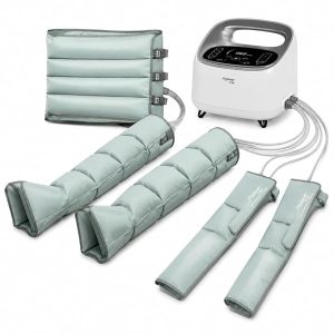
Limb Compression Therapy System

Limb Compression Therapy System

Limb Compression Therapy System: Advanced Support for Circulation, Recovery, and Patient Care

Poor circulation, swelling, and reduced mobility can significantly affect a patient’s comfort and recovery. A Limb Compression Therapy System offers a safe, non-invasive solution by applying controlled pressure to the arms or legs. This therapy helps improve blood circulation, support venous return, reduce fluid accumulation, and enhance patient comfort.

Modern limb compression systems are increasingly used in hospitals, clinics, rehabilitation centers, physiotherapy departments, vascular practices, and home-care settings.Limb Compression Therapy System

What Is a Limb Compression Therapy System?

A Limb Compression Therapy System—also known as an Intermittent Pneumatic Compression (IPC) system—typically consists of a programmable compression pump and inflatable sleeves or garments.

The sleeves are placed around the patient’s leg, foot, or arm. Air is then delivered into different chambers of the garment in a controlled sequence. These chambers inflate and deflate rhythmically, creating a gentle compression cycle that supports the natural movement of blood and lymphatic fluid.

Depending on the device and clinical purpose, the healthcare professional may adjust:

  • Compression pressure
  • Treatment duration
  • Inflation and deflation cycles
  • Compression mode
  • Chamber sequence
  • Treatment area

How Does Compression Therapy Work?

When limb movement is limited, the natural muscle-pump mechanism becomes less effective. Blood and fluid may accumulate in the extremities, causing swelling, discomfort, and circulatory complications.

A pneumatic compression system simulates the effect of muscle movement by applying sequential external pressure. This may help:

  • Encourage venous blood to return toward the heart
  • Reduce venous stasis
  • Support lymphatic drainage
  • Decrease limb swelling
  • Improve local circulation
  • Reduce sensations of heaviness and discomfort

Sequential compression usually begins at the most distant part of the limb and progresses upward. This graduated action helps move accumulated blood and fluid in the appropriate direction.

Clinical Applications

1. Deep Vein Thrombosis Prevention

Patients who remain immobile for extended periods may have an increased risk of developing deep vein thrombosis (DVT). This is particularly relevant for hospitalized patients and those recovering after major surgery.

When prescribed appropriately, intermittent pneumatic compression may be included as part of a comprehensive DVT-prevention protocol. Clinical assessment remains essential, as some patients may also require medication or additional preventive measures.

2. Postoperative Patient Care

Following surgery, pain, weakness, or restricted movement can reduce normal lower-limb activity. A limb compression system may support venous circulation during the recovery period.

It may be considered for patients following:

  • Orthopedic surgery
  • General surgery
  • Gynecological surgery
  • Bariatric surgery
  • Neurological procedures
  • Other major operations associated with reduced mobility

3. Lymphedema Management

Lymphedema occurs when lymphatic fluid accumulates in the tissues, producing persistent swelling. Compression therapy may be used as one component of a broader lymphedema-management program.

Depending on the patient’s condition, the complete treatment plan may also include manual lymphatic drainage, exercise, skincare, compression garments, and specialist follow-up.

4. Management of Limb Edema

Swelling of the arms or legs can result from several clinical conditions. When the underlying cause has been evaluated and compression is considered suitable, pneumatic compression may help reduce fluid accumulation and improve comfort.

Healthcare professionals should always investigate the cause of unexplained or sudden swelling before initiating treatment.

5. Rehabilitation and Physiotherapy

Patients recovering from injury, illness, or prolonged immobilization may benefit from compression therapy as part of a supervised rehabilitation program. Improved circulation and reduced swelling may help patients participate more comfortably in movement and therapeutic exercise.

6. Vascular and Venous Care

Limb compression systems may support selected patients with venous circulation problems. However, treatment pressure and duration should be determined carefully, particularly in patients with arterial disease, diabetes, neuropathy, fragile skin, or complex vascular conditions.

Benefits of a Modern Limb Compression Therapy System

A well-designed compression therapy system can provide several clinical and operational advantages:

  • Non-invasive treatment
  • Adjustable compression pressure
  • Consistent and repeatable therapy cycles
  • Sequential or graduated compression
  • Simple application and operation
  • Multiple sleeve or garment options
  • Improved patient comfort
  • Support for hospital and home-care workflows
  • Reduced manual effort for healthcare staff
  • Potential support for faster rehabilitation

Advanced models may also include digital controls, real-time pressure monitoring, multiple treatment programs, alarms, and patient-specific settings.

Departments That May Use Limb Compression Therapy

Limb compression systems can support patient care across multiple medical specialties, including:

  • Vascular surgery
  • Orthopedics
  • General surgery
  • Anesthesiology
  • Intensive care
  • Physical medicine and rehabilitation
  • Physiotherapy
  • Oncology
  • Neurology
  • Gynecology and obstetrics
  • Lymphedema clinics
  • Postoperative recovery units
  • Long-term and home care

Patient Safety and Contraindications

Compression therapy is not appropriate for every patient. A qualified healthcare professional should evaluate the patient before treatment begins.

Extra caution or avoidance may be necessary in cases such as:

  • Known or suspected acute DVT
  • Severe peripheral arterial disease
  • Acute thrombophlebitis
  • Decompensated heart failure
  • Severe limb infection or cellulitis
  • Open wounds without appropriate protection
  • Severe neuropathy or loss of sensation
  • Unexplained pain or sudden swelling
  • Conditions where increased venous or lymphatic return may be harmful

The patient’s circulation, skin condition, sensation, medical history, and treatment response should be monitored throughout therapy. The device must always be used according to the manufacturer’s instructions and the clinician’s prescription.

What to Consider When Selecting a System

Healthcare facilities should consider both clinical performance and long-term usability when choosing a limb compression therapy system.

Important factors include:

  • Accurate and adjustable pressure control
  • Sequential compression capability
  • Number and arrangement of air chambers
  • Range of available sleeve sizes
  • Upper- and lower-limb compatibility
  • Easy-to-clean materials
  • Clear display and user-friendly controls
  • Safety alarms
  • Portability
  • Noise level
  • Durability
  • Availability of accessories
  • Warranty and technical support
  • User training and after-sales service

The most suitable system is not simply the one with the highest pressure. It is the system that provides reliable, controlled, comfortable, and clinically appropriate compression for the intended patient group.

Limb Compression Therapy Solutions from Unique Medi Trade

Unique Medi Trade is committed to introducing advanced medical technologies that support safer treatment, efficient clinical workflows, and improved patient care in Bangladesh.

We help healthcare providers select suitable Limb Compression Therapy Systems for hospitals, surgical units, rehabilitation centers, physiotherapy facilities, vascular practices, and home-care services. Our support may include product consultation, demonstrations, user training, installation guidance, and dependable after-sales service.

Conclusion

A Limb Compression Therapy System can play an important role in circulation support, edema management, rehabilitation, and postoperative care. Through controlled and sequential pneumatic pressure, it helps support venous return and fluid movement while providing a convenient, non-invasive treatment option.

Successful therapy depends on selecting the right system, assessing the patient properly, using appropriate settings, and following established clinical protocols.

Looking for a reliable Limb Compression Therapy System in Bangladesh? Contact Unique Medi Trade for product information, clinical demonstrations, pricing, and professional support.

Contact: +8801717811312

Medical disclaimer: This article is intended for educational purposes only. Limb compression therapy should be prescribed and supervised by a qualified healthcare professional.

POINT-OF-CARE ULTRASOUND IN PAIN MEDICINE

Point-of-Care Ultrasound (POCUS) in Vascular Medicine & Vascular Surgery

Point-of-Care Ultrasound (POCUS) in Vascular Medicine and Vascular Surgery: See the Vessel, Understand the Flow, Guide the Procedure

Bringing Vascular Ultrasound Directly to the Point of Care

Vascular medicine is a specialty built around anatomy, circulation and blood flow. A patient may present with: Leg swelling.Point-of-Care Ultrasound (POCUS) in Vascular Medicine & Vascular Surgery Sudden limb pain. Varicose veins. Suspected Deep Vein Thrombosis (DVT). Poor peripheral circulation. A difficult vascular access. An arteriovenous fistula problem. Or a vascular surgeon may need to perform an ultrasound-guided endovenous intervention. In all of these situations, seeing the vessel can provide valuable information that physical examination alone cannot provide. This is where Point-of-Care Ultrasound (POCUS) is becoming increasingly relevant. POCUS allows an appropriately trained physician to perform a focused ultrasound examination at the bedside, outpatient chamber, procedure room or operating environment and immediately integrate the findings into clinical decision-making. A recent review of vascular POCUS describes its expanding use beyond procedural guidance, particularly for evaluation of lower-extremity deep venous thrombosis and the abdominal aorta. For vascular specialists, however, its potential goes even further.
Vein. Artery. Flow. Needle. Catheter.

POCUS puts vascular anatomy directly into the physician's hands.


What Is Vascular POCUS?

Point-of-Care Ultrasound is generally a focused examination performed and interpreted by the treating clinician to answer a specific clinical question. For a vascular specialist, those questions may include:
  • Is this vein compressible? Point-of-Care Ultrasound (POCUS) in Internal Medicine
  • Is proximal DVT suspected?
  • Where is the target vein?
  • Where is the artery?
  • What is the diameter and depth of the vessel?
  • Which vessel is most appropriate for access?
  • Is blood flow visible with Color Doppler?
  • Is there a superficial venous abnormality?
  • Is the abdominal aorta enlarged?
  • Is an AV fistula accessible for cannulation?
  • Is there a collection, hematoma or suspected pseudoaneurysm?
  • Where should an endovenous catheter or needle be positioned?
  • What structures should be avoided during an intervention?
This question-focused approach is different from a complete vascular laboratory duplex examination. POCUS is designed primarily to provide immediate, targeted information.

Why POCUS Is Particularly Relevant to Vascular Specialists

The vascular specialist works with structures that are highly suitable for ultrasound. Many important vessels are relatively superficial and can be clearly differentiated using B-mode and Doppler imaging. With ultrasound, the physician can potentially evaluate: 

Vessel Anatomy

Location, depth, diameter and relationship with surrounding structures.

Compressibility

Particularly relevant in venous thrombosis assessment.

Blood Flow

Using Color and spectral Doppler when appropriate.

Needle Position

During vascular access and other interventions.

Surrounding Anatomy

Including nerves, muscles, soft tissue and adjacent arteries or veins. This creates an important transition in vascular practice:

From feeling the pulse to seeing the artery.

From locating the vein by landmarks to visualizing it.

From estimating needle position to guiding it in real time.


1. Point-of-Care Ultrasound for Deep Vein Thrombosis

Deep Vein Thrombosis is one of the most established diagnostic applications of vascular POCUS. Patients may present with:
  • Unilateral leg swelling Point-of-Care Ultrasound (POCUS) in Vascular Medicine & Vascular Surgery
  • Leg pain
  • Tenderness
  • Increased limb circumference
  • Risk factors for venous thromboembolism
Clinical examination alone cannot reliably confirm or exclude DVT. Focused compression ultrasound can allow a trained physician to assess whether key proximal veins compress normally. The fundamental concept is simple:

Normal vein → Usually compresses under appropriate probe pressure.

Thrombosed vein → May fail to compress normally.

A 2024 systematic review and meta-analysis found strong diagnostic performance for several clinician-performed POCUS protocols for DVT, although performance depends on technique, training and the specific protocol used. A dedicated review also concluded that POCUS can have excellent diagnostic accuracy for acute proximal lower-extremity DVT when performed by well-trained users, while emphasizing the need for appropriate education and standardized training. For a vascular physician evaluating a patient with suspected DVT, this means bedside ultrasound may provide clinically important information immediately.

2. Compression Ultrasound: A Practical Vascular POCUS Skill

Compression ultrasound is one of the fundamental skills of venous POCUS. The physician identifies the artery and vein in transverse view and applies controlled pressure with the probe. The examination may assess selected venous segments according to the protocol being used. A vascular POCUS examination may incorporate: B-Mode for anatomy and compressibility, Color Doppler for visualization of flow, and when appropriate, Spectral Doppler for additional hemodynamic information. Professional ultrasound guidance for peripheral venous examinations emphasizes structured assessment and documentation for complete diagnostic studies. This highlights an important distinction:

Focused DVT POCUS and comprehensive venous duplex ultrasound are related—but they are not always interchangeable.

If findings are uncertain, symptoms suggest distal or complex disease, or detailed venous mapping is required, a complete vascular duplex examination may still be necessary.

3. Varicose Veins and Chronic Venous Disease

Chronic venous disease is a major part of vascular practice. Patients may present with:
  • Varicose veins  Point-of-Care Ultrasound (POCUS) in Vascular Medicine & Vascular Surgery
  • Leg heaviness
  • Pain
  • Swelling
  • Skin pigmentation
  • Venous eczema
  • Lipodermatosclerosis
  • Venous ulceration
Ultrasound plays a fundamental role in evaluating venous anatomy and reflux. Current Society for Vascular Surgery, American Venous Forum and American Vein and Lymphatic Society guidelines include duplex ultrasound scanning as a central component in evaluating patients with lower-extremity varicose veins and superficial truncal reflux. For the vascular specialist, a handheld ultrasound can be useful during the initial clinical encounter for focused questions. For example: Which superficial vein is visible beneath the symptomatic area? Is the vessel patent? What is its approximate diameter and depth? Is obvious reflux or abnormal flow suspected? However, treatment planning for chronic venous insufficiency generally requires a structured duplex study, including detailed reflux assessment and mapping. POCUS can complement that workflow rather than replace it.

4. Ultrasound-Guided Endovenous Radiofrequency Ablation

One of the strongest examples of ultrasound becoming an integral part of vascular intervention is Endovenous Radiofrequency Ablation (RFA). Endovenous thermal ablation is used in appropriately selected patients with superficial venous reflux and varicose vein disease. During a typical ultrasound-guided procedure, ultrasound may assist the physician in:

Identifying the Target Vein Point-of-Care Ultrasound (POCUS) in Vascular Medicine & Vascular Surgery

Selecting an Access Site

Guiding Needle Entry

Guiding Guidewire and Catheter Position

Evaluating the Relationship to Deep Veins

Guiding Tumescent Anaesthesia

Monitoring the Procedure

Assessing the Treated Vein After Intervention

International consensus describes endovenous thermal ablation—including radiofrequency—as a catheter-directed, ultrasound-guided treatment for varicose vein disease. A multidisciplinary international position statement also recommends concurrent ultrasound imaging for superficial endovenous procedures including radiofrequency ablation, endovenous laser ablation, cyanoacrylate closure and ultrasound-guided sclerotherapy. This makes ultrasound much more than a diagnostic tool for vascular surgeons.

It becomes a procedural navigation tool.


5. Ultrasound-Guided Vascular Access

One of the most practical uses of POCUS is vascular access. Every clinician knows the challenge: A vein may be palpable in one patient but almost impossible to identify in another. Ultrasound allows the operator to visualize: Vein Artery Depth Diameter Surrounding structures Needle before and during cannulation. POCUS can be used for:
  • Peripheral venous access
  • Central venous access
  • Selected arterial access
  • Difficult vascular access
  • Dialysis access-related procedures
International evidence-based recommendations support ultrasound guidance for central venous cannulation and note its value in selected peripheral venous and arterial access procedures. A review of Cochrane evidence found ultrasound-guided vascular access associated with improvements in outcomes such as first-attempt success and overall success in several studied settings, while also reducing some failed attempts and complications.

6. Seeing the Needle During Vascular Access

A major advantage of ultrasound guidance is the ability to observe needle advancement. Two commonly used techniques are:

In-Plane

The needle advances along the ultrasound imaging plane, potentially allowing visualization of much of the needle shaft and tip.

Out-of-Plane

The needle crosses the ultrasound beam and appears as a bright echogenic point when intersecting the imaging plane. The important principle is:

Always understand where the needle tip is.

Seeing part of the needle does not necessarily mean the operator has identified the tip. Training in hand-eye coordination, probe positioning and needle visualization is therefore essential.

7. Arterial POCUS

Arterial ultrasound can provide valuable focused information in vascular practice. Depending on the clinical situation and device capability, a vascular physician may use ultrasound to evaluate:
  • Arterial location
  • Vessel diameter
  • Patency
  • Pulsatility
  • Calcification
  • Selected flow characteristics
  • Suitable arterial access sites
  • Surrounding anatomy
Color Doppler can help differentiate arterial and venous flow, while spectral Doppler may provide additional hemodynamic information. For comprehensive assessment of Peripheral Arterial Disease (PAD), however, formal vascular testing may involve:
  • Ankle-Brachial Index
  • Segmental pressures
  • Pulse-volume recordings
  • Complete arterial duplex
  • CT angiography
  • MR angiography
  • Conventional angiography
depending on the clinical situation. Therefore:

POCUS can answer focused arterial questions, but it should not automatically be considered a substitute for comprehensive vascular investigation.


8. Abdominal Aortic Aneurysm

The abdominal aorta is another important target for POCUS. A focused transabdominal examination can help determine whether the aorta appears enlarged in a patient where an abdominal aortic aneurysm is suspected. This may be particularly relevant for patients presenting with combinations such as: Abdominal pain Back pain Hypotension Syncope and appropriate vascular risk factors. Recent evidence continues to support the ability of trained clinicians to detect AAA with POCUS. A 2026 review of systematic reviews reported very high ranges of sensitivity and specificity for clinician-performed POCUS in diagnosing AAA. A 2024 systematic review and meta-analysis similarly reported high diagnostic performance in emergency settings. For a vascular specialist, however, identifying an aneurysm is only the beginning. Detailed treatment planning may require comprehensive ultrasound, CT angiography or other vascular imaging to assess anatomy, branch vessels, morphology and suitability for intervention.

9. Pseudoaneurysm Assessment

Pseudoaneurysm can occur following arterial puncture, catheterization, trauma or vascular procedures. Ultrasound can help evaluate a suspicious groin or access-site swelling and identify vascular flow patterns suggesting a pseudoaneurysm. Depending on the case and operator expertise, ultrasound may also support selected image-guided treatment. EFSUMB guidance on ultrasound-guided vascular interventions includes ultrasound for detection of vascular-access complications and ultrasound-guided treatment of arterial pseudoaneurysms. This is another example of POCUS moving beyond simple diagnosis into intervention.

10. Arteriovenous Fistula and Dialysis Access

Patients receiving haemodialysis depend on reliable vascular access. POCUS can be used for focused evaluation of an AV fistula or graft. The physician may assess questions such as:
  • Where is the fistula?
  • How deep is the vessel?
  • What is its diameter?
  • Is there visible flow?
  • Is there an obvious abnormality?
  • Where is the most appropriate area for cannulation?
A review of vascular-access POCUS emphasizes that its goal is often to answer focused bedside questions rather than reproduce a complete fistula or graft duplex examination. A broader review found POCUS has been used for AV fistula maturation assessment, identification of abnormalities, difficult cannulation and selection of alternative cannulation sites. This distinction perfectly illustrates the philosophy of POCUS:

Ask a focused question.

Scan.

Obtain immediate information.

Decide whether additional investigation is required.


11. Color Doppler: Seeing More Than Anatomy

B-mode ultrasound shows anatomy. Color Doppler adds information about blood flow. For vascular physicians, this can be particularly valuable. Color Doppler may help identify:
  • Vessel flow
  • Flow direction
  • Areas of abnormal flow
  • Relationship between vessels
  • Possible vascular abnormalities
  • Patency during focused assessment
Spectral Doppler can further provide information about flow velocity and waveform morphology when the equipment and operator training support it. For advanced vascular assessment, Doppler capability becomes especially important because vascular medicine is not simply about seeing vessels.

It is about understanding circulation.


12. POCUS Before a Vascular Procedure

Imagine preparing for an intervention. Before puncturing the skin, the physician can scan the area and ask:

Where exactly is the artery?

Where is the vein?

How deep is the vessel?

What is its diameter?

Is there another vessel nearby?

Is there calcification?

Is this the best access point?

What is the safest needle trajectory?

This brief pre-procedural scan can transform a landmark-based procedure into an image-informed procedure. International vascular-access recommendations support ultrasound screening before cannulation and real-time needle guidance in appropriate settings.

13. POCUS During the Procedure

The value continues after skin puncture. Real-time ultrasound can potentially allow the operator to see: Needle → Vessel → Guidewire → Catheter depending on the procedure. For endovenous treatment, ultrasound may also be used to observe surrounding anatomy and assist with proper treatment-zone positioning. This creates a major difference between:

“I believe the device is in the correct location.”

and

“I can visualize where it is.”


14. POCUS After the Procedure

Focused ultrasound may also contribute to immediate post-procedure assessment. Depending on the intervention, the physician may look for:
  • Vessel patency
  • Treatment response
  • Hematoma
  • Suspected access-site complication
  • Pseudoaneurysm
  • Local fluid collection
  • Other relevant findings
More comprehensive post-treatment surveillance may still require a formal vascular duplex study according to the procedure and clinical guideline. Duplex ultrasound remains a fundamental part of assessment following many venous interventions.

POCUS and Endovenous RFA: A Natural Combination

For a vascular practice performing Radiofrequency Ablation for Varicose Veins, ultrasound and RFA naturally work together. A simplified clinical workflow may look like: Clinical AssessmentDuplex EvaluationTreatment PlanningUltrasound-Guided Venous AccessCatheter PositioningTumescent Anaesthesia Under Ultrasound GuidanceRadiofrequency AblationUltrasound Assessment Ultrasound provides visualization. Radiofrequency provides the therapeutic energy. Together, these technologies support modern minimally invasive treatment of appropriately selected superficial venous disease.

Which Ultrasound Probe Does a Vascular Specialist Need?

Linear Probe — The Essential Vascular Probe

For most superficial vascular applications, a high-frequency Linear probe is particularly important. It is useful for:
  • DVT assessment
  • Superficial veins
  • Vascular access
  • Carotid and other superficial vessels
  • Varicose veins
  • Endovenous procedures
  • AV fistula
  • Peripheral arteries
  • Needle guidance
  • Soft-tissue assessment
The rectangular image and high-frequency capability make the linear transducer particularly suitable for superficial anatomy.

Why a Convex Probe Can Also Be Useful

Not every vessel is superficial. For deeper structures, the Convex probe can provide greater penetration and a wider field of view. Potential applications include:
  • Abdominal aorta
  • Iliac-region assessment
  • Deep abdominal structures
  • Selected deep vascular anatomy
  • Abdominal POCUS
This means that a vascular specialist may benefit from having:

Linear for superficial vascular imaging

and

Convex for deeper abdominal vascular assessment.


Why Handheld Wireless Ultrasound Makes Sense in Vascular Practice

Traditional vascular ultrasound systems remain essential for detailed duplex examinations. But a vascular surgeon does not perform every clinical assessment inside a vascular laboratory. The specialist may be working in:
  • Consultation chamber
  • Vascular clinic
  • Hospital ward
  • Emergency department
  • Procedure room
  • Operating theatre
  • Dialysis unit
  • Bedside
  • Peripheral healthcare facility
A handheld wireless ultrasound can travel with the physician. It can potentially connect to a compatible smartphone or tablet and provide immediate imaging when a focused clinical question arises. The workflow becomes:

Examine → Scan → Visualize → Decide → Intervene when appropriate.

This is the philosophy behind Point-of-Care Ultrasound.

SonoHealth Handheld Wireless Ultrasound for Vascular POCUS

For vascular specialists looking to introduce portable ultrasound into everyday practice, SonoHealth Handheld Wireless Ultrasound provides a practical point-of-care imaging platform. Depending on the configuration, it can support:

Vascular Imaging

Focused examination of veins and arteries.

Color Doppler

Visualization of vascular flow.

DVT Assessment

Focused compression ultrasound in appropriately selected patients.

Vascular Access

Real-time ultrasound guidance for needle placement.

Endovenous Procedures

Support for image-guided vascular intervention.

AV Fistula Assessment

Focused evaluation and access guidance.

Abdominal Vascular Assessment

With an appropriate deeper-penetration transducer.

Needle Guidance

For selected vascular procedures. For many vascular applications, the combination of a high-frequency Linear probe + Color Doppler + needle-guidance capability can be particularly useful. Adding a Convex probe expands the potential range toward deeper abdominal and aortic imaging.

POCUS Does Not Replace a Complete Vascular Duplex Study

This is one of the most important principles when adopting handheld ultrasound. A focused POCUS examination and a comprehensive vascular duplex examination are not necessarily the same thing. A complete vascular laboratory examination may require:
  • Detailed vessel mapping
  • Standardized Doppler waveforms
  • Velocity measurements
  • Reflux testing
  • Spectral Doppler analysis
  • Multiple standardized anatomical segments
  • Formal interpretation
  • Structured reporting
  • Quality-control protocols
Professional vascular guidelines continue to rely heavily on comprehensive duplex ultrasound for conditions such as chronic venous disease and treatment planning. Therefore, POCUS should be considered:

A focused clinical tool.

A procedural guidance tool.

An extension of vascular examination.

not automatically a replacement for comprehensive vascular imaging.

Training Matters

Ultrasound is operator-dependent. A sophisticated ultrasound device is only useful when the physician understands what is being displayed. Vascular POCUS training should include:
  • Ultrasound physics
  • Probe orientation
  • Vascular anatomy
  • Artery-versus-vein identification
  • Compression technique
  • B-mode optimization
  • Color Doppler
  • Spectral Doppler where applicable
  • Needle visualization
  • In-plane and out-of-plane approaches
  • Recognition of artifacts
  • Recognition of DVT findings
  • Recognition of vascular-access complications
  • Understanding when comprehensive imaging is required
Published evidence on DVT POCUS repeatedly emphasizes that good diagnostic performance depends on adequately trained operators.

Technology + Training + Clinical Knowledge = Effective Vascular POCUS.


The Future of Vascular Medicine Is Increasingly Visual

Vascular specialists have always relied heavily on clinical examination. They inspect the limb. They palpate pulses. They evaluate edema. They assess skin changes. They listen for bruits. POCUS adds another dimension:

They can see the vessel.

They can see flow.

They can see the needle.

They can see the target before intervention.

This does not replace clinical examination. It strengthens it.

From Palpation to Visualization

A handheld ultrasound can change the everyday vascular workflow. Instead of: “I think the vein is here.” the physician may be able to say:

“I can see the vein.”

Instead of: “The vessel should be approximately this deep.”

“I can measure its depth.”

Instead of: “The needle should be approaching the vessel.”

“I can follow the needle toward the target.”

Instead of relying solely on external anatomy:

The vascular specialist can visualize what lies underneath.

That is the practical value of POCUS.

See the Vessel. Understand the Flow. Guide the Procedure.

Point-of-Care Ultrasound is helping bring real-time vascular imaging closer to the physician and closer to the patient. For vascular specialists, its potential spans: DVT Assessment Venous Examination Arterial Assessment Vascular Access Varicose Vein Procedures Endovenous RFA AV Fistula Assessment Aortic POCUS Needle Guidance and selected post-procedural assessments. The future is not simply about smaller ultrasound machines. It is about having relevant imaging available when the clinical decision is being made.

SonoHealth Handheld Wireless Ultrasound

Point-of-Care Ultrasound for Modern Vascular Practice

Vascular Imaging • Color Doppler • DVT Assessment • Vascular Access • Endovenous Procedures • Needle Guidance

See the Vessel. Understand the Flow. Guide with Precision.

For vascular surgeons, vascular specialists, hospitals and vascular centers interested in integrating handheld Point-of-Care Ultrasound into clinical practice: Unique Medi Trade POCUS Solutions • Live Demonstration • Clinical Training Support • Technical Support • Endovenous RFA Solutions Contact: +880 1717-811312 Unique Medi Trade — Advancing Image-Guided Vascular Care in Bangladesh

Medical Disclaimer

This article is intended for healthcare-professional education and general information. Point-of-Care Ultrasound should be performed by appropriately trained healthcare professionals within their scope of practice and according to relevant clinical guidelines and institutional protocols. Focused POCUS should not replace a comprehensive vascular duplex examination, CT angiography, MR angiography, conventional angiography or specialist vascular imaging when these are clinically indicated. Clinical decisions should be based on the complete patient assessment rather than an isolated POCUS finding.

SEO Information

SEO Title: Point-of-Care Ultrasound (POCUS) in Vascular Medicine & Vascular Surgery Alternative SEO Title: POCUS for Vascular Surgeons: DVT, Vascular Access, Varicose Veins & Endovenous Procedures Meta Description: Discover how Point-of-Care Ultrasound (POCUS) supports vascular specialists in DVT assessment, vascular access, venous and arterial imaging, endovenous RFA, AV fistula assessment and image-guided procedures. Suggested URL Slug: /point-of-care-ultrasound-pocus-in-vascular-surgery/ Target Keywords: Point-of-Care Ultrasound Vascular, POCUS Vascular Surgery, vascular ultrasound, handheld vascular ultrasound, DVT ultrasound, ultrasound-guided vascular access, varicose vein ultrasound, Endovenous RFA, Color Doppler ultrasound, vascular surgeon ultrasound, SonoHealth Handheld Ultrasound, POCUS Bangladesh Suggested Featured Image Text: POINT-OF-CARE ULTRASOUND IN VASCULAR MEDICINE See the Vessel. Understand the Flow. Guide with Precision. SonoHealth Handheld Wireless Ultrasound

POINT-OF-CARE ULTRASOUND IN PAIN MEDICINE

Point-of-Care Ultrasound (POCUS) in Pain Medicine

Point-of-Care Ultrasound (POCUS) in Pain Medicine: Seeing the Target, Guiding the Needle, Improving Precision

A New Dimension in Modern Pain Management

Pain Medicine is becoming increasingly precise, image-guided and minimally invasive. Traditionally, many pain procedures were performed using anatomical landmarks, palpation, nerve stimulation, fluoroscopy or CT guidance. Today, Point-of-Care Ultrasound (POCUS) has added another powerful option to the pain physician's toolkit. With ultrasound available directly at the point of care, an appropriately trained pain physician can visualize important structures in real time—including: Nerves • Muscles • Fascia • Tendons • Joints • Blood Vessels • Pleura • Bone Surfaces • Needle Path This ability to see anatomy while performing an intervention can fundamentally change the way selected pain procedures are planned and performed. The American Society of Regional Anesthesia and Pain Medicine (ASRA) describes POCUS as an important skill for regional anesthesiologists and pain physicians and has published expert recommendations regarding its clinical indications and training. For the modern pain specialist, ultrasound is therefore becoming more than an imaging machine.

It can become a visual extension of the clinical examination—and a real-time navigation system for interventions.


What Is Point-of-Care Ultrasound in Pain Medicine?

Point-of-Care Ultrasound means performing a focused ultrasound examination at or near the patient's bedside, consultation room or procedure area to answer a specific clinical question or guide an intervention. In Pain Medicine, the physician may ask:Point-of-Care Ultrasound (POCUS) in Internal Medicine Where exactly is the target nerve? Where is the needle tip? Are there blood vessels close to my planned needle path? How deep is the target? Is the injectate spreading around the intended structure? Is there a joint effusion? Can I identify the muscle, tendon or fascial plane responsible for the patient's symptoms? Rather than relying only on surface anatomy, ultrasound allows the physician to visualize structures beneath the skin in real time. That is one of the major reasons ultrasound guidance has become increasingly important in regional anesthesia and interventional pain practice. Research and expert reviews describe applications ranging from peripheral nerve blocks and fascial-plane procedures to musculoskeletal and chronic pain interventions.

From Landmark-Guided to Image-Guided Pain Intervention

Consider a traditional injection performed primarily using anatomical landmarks. The physician knows where the nerve or joint should normally be located based on anatomy. But every patient is different. Body habitus varies. Muscle thickness varies. Nerve location may vary. Blood vessels may be closer to the intended needle trajectory than expected. Scar tissue or previous surgery may alter anatomy. Ultrasound changes this approach. Instead of thinking:

“The target should be here.”

The physician may be able to say:

“I can see the target here.”

And instead of advancing the needle based primarily on anatomical estimation:

The physician can potentially visualize the needle approaching the target in real time.

This is the fundamental advantage of ultrasound-guided pain intervention.

Why Ultrasound Is Particularly Valuable in Pain Medicine

Many pain procedures involve relatively small anatomical targets surrounded by structures that physicians would prefer to avoid. For example:POINT-OF-CARE ULTRASOUND IN PAIN MEDICINE Target nerve may be located beside a: Blood vessel or beneath a: Muscle or fascial layer or near: Pleura or another sensitive structure. Ultrasound provides real-time anatomical information that can help the trained operator plan a more informed needle trajectory. A review of ultrasound-guided chronic pain interventions highlights the ability of ultrasound to visualize the target tissue, surrounding vascular structures and distribution of injected medication. This is particularly relevant in procedures where millimeters matter.

1. Peripheral Nerve Blocks

Peripheral nerve blocks are among the most important applications of ultrasound in pain medicine. Using a high-frequency linear probe, physicians can often identify superficial peripheral nerves and surrounding anatomical structures. Depending on indication, training and scope of practice, ultrasound-guided interventions may involve nerves such as:
  • Suprascapular nerve
  • Median nerve
  • Ulnar nerve
  • Radial nerve
  • Greater occipital nerve
  • Lateral femoral cutaneous nerve
  • Ilioinguinal nerve
  • Iliohypogastric nerve
  • Genitofemoral nerve
  • Genicular nerves
  • Selected ankle and foot nerves
  • Other peripheral nerve targets
A contemporary review of chronic pain interventions describes ultrasound-guided techniques involving many of these peripheral nerve targets. The physician can potentially visualize: Nerve → Needle → Adjacent vessels → Surrounding tissue → Injectate spread during the intervention.

2. Ultrasound-Guided Diagnostic Nerve Blocks

Pain Medicine frequently involves answering an important question:

“Is this structure actually responsible for the patient's pain?”

A diagnostic block can sometimes help answer that question. For selected conditions, a physician may inject a local anesthetic around a suspected nerve or anatomical target and evaluate the patient's response. Ultrasound can assist with accurate localization of selected peripheral targets.POINT-OF-CARE ULTRASOUND IN PAIN MEDICINE This can be valuable because interventional pain management is not simply about treating pain. It is also about identifying the pain generator as accurately as possible. A carefully performed diagnostic block may therefore become part of the clinical decision-making process before considering longer-lasting interventions.

3. Ultrasound and Radiofrequency Procedures

Radiofrequency technology plays an important role in modern interventional Pain Medicine. In selected procedures, a physician first identifies the relevant nerve or target structure and then positions a radiofrequency electrode before delivering controlled RF energy. Depending on the anatomical target, procedure and physician expertise, ultrasound may assist with target identification and needle/electrode placement. Published reviews of ultrasound-guided chronic pain procedures include both diagnostic nerve blocks and radiofrequency-related interventions. This creates an important relationship between two technologies:

Ultrasound helps the physician see.

Radiofrequency helps the physician treat selected pain targets.

For a pain practice developing advanced interventional services, these technologies can therefore complement each other in appropriately selected procedures. However, not every radiofrequency procedure should be performed using ultrasound alone. Fluoroscopy, CT or other guidance may remain preferred or necessary for certain spinal and deep anatomical targets.

4. Shoulder Pain

Shoulder pain is one of the most common musculoskeletal complaints encountered in clinical practice. Several structures may potentially contribute to shoulder pain, including:
  • Rotator cuff tendonsPOINT-OF-CARE ULTRASOUND IN PAIN MEDICINE
  • Subacromial-subdeltoid bursa
  • Biceps tendon
  • Acromioclavicular joint
  • Glenohumeral joint
  • Suprascapular nerve
  • Surrounding muscles and soft tissues
Musculoskeletal ultrasound allows the physician to dynamically assess many superficial shoulder structures. For appropriately selected interventions, ultrasound can also guide the needle toward a specific target. One important pain intervention is the suprascapular nerve block, which has applications in selected chronic shoulder pain conditions. Ultrasound-guided procedures targeting the suprascapular nerve are among techniques described in reviews of chronic pain intervention.

5. Knee Pain

Chronic knee pain can result from multiple conditions, including osteoarthritis and other musculoskeletal disorders. Ultrasound may assist the physician in evaluating:
  • Joint effusion
  • Superficial tendons
  • Selected ligaments
  • Periarticular soft tissues
  • Baker's cyst
  • Selected peripheral nerve targets
Ultrasound can also guide selected knee injections. Another increasingly important interventional target in chronic knee pain is the genicular nerve system. Ultrasound-guided genicular nerve interventions, including blocks and radiofrequency-related techniques, are described in the pain literature. (PubMed) For a physician treating chronic knee pain, ultrasound therefore has potential value across both assessment and intervention.

6. Hip Pain

The hip is anatomically deeper than many other musculoskeletal targets, making appropriate probe selection and operator training especially important. Ultrasound-guided pain procedures around the hip have continued to develop, and contemporary literature describes ultrasound applications for chronic pain involving the hip, shoulder, knee and lumbar regions. (PubMed) Depending on the clinical condition, ultrasound may assist in visualization of selected:
  • TendonsPOINT-OF-CARE ULTRASOUND IN PAIN MEDICINE
  • Muscles
  • Bursae
  • Joint-related structures
  • Peripheral nerves
For deeper targets, a lower-frequency or convex probe may sometimes be more suitable than a high-frequency linear probe. This is why probe selection matters in Pain Medicine.

7. Myofascial Pain and Trigger-Point Procedures

Myofascial pain is frequently encountered in Pain Medicine. Patients may present with:
  • Localized muscle pain
  • Trigger points
  • Referred pain
  • Muscle spasm
  • Functional limitation
Trigger-point injections have traditionally often been performed using palpation. Ultrasound can provide additional information regarding muscle layers and surrounding anatomy, particularly for deeper targets. A systematic review of ultrasound-guided interventions for myofascial trigger points found some evidence supporting ultrasound guidance over blinded interventions, while also emphasizing that the available studies had limitations and further high-quality research is needed. (PubMed) This is an important principle in responsible POCUS practice:

Ultrasound can improve visualization, but clinical outcomes depend on much more than imaging alone.

Correct diagnosis, patient selection, technique and appropriate treatment strategy remain essential.

8. Fascial Plane Blocks

Modern ultrasound has contributed significantly to the development of fascial-plane blocks. Rather than always targeting one clearly defined peripheral nerve, the physician may place medication within an anatomical fascial plane where relevant nerves travel. Examples described in pain and regional anesthesia literature include:
  • Transversus Abdominis Plane (TAP) blockPOINT-OF-CARE ULTRASOUND IN PAIN MEDICINE
  • Quadratus Lumborum block
  • Rectus sheath block
  • Erector Spinae Plane block
  • Pectoral plane blocks
  • Serratus plane blocks
Ultrasound allows physicians to identify muscle and fascial layers and observe the spread of injectate within the intended plane. Reviews of ultrasound-guided chronic pain interventions include many of these fascial-plane techniques. (PubMed)

9. Occipital Neuralgia and Headache-Related Pain

The greater occipital nerve is another potential target in selected pain conditions. For patients with appropriate headache syndromes or occipital neuralgia, an ultrasound-guided approach may help the physician identify the relevant anatomical region and nearby structures. The greater occipital nerve is among the targets covered in current reviews of ultrasound-guided chronic pain procedures. (PubMed) Again, ultrasound does not determine the diagnosis by itself. It supports a procedure after the clinician has established an appropriate indication based on history, examination and clinical evaluation.

10. Ultrasound-Guided Joint Injections

Joint injections are commonly used in musculoskeletal and pain practice. Depending on the condition and physician expertise, ultrasound may be used to guide injections involving selected: Shoulder jointsPOINT-OF-CARE ULTRASOUND IN PAIN MEDICINE Knee-related structures Hip-related structures Small peripheral joints and periarticular targets. The value of imaging guidance is straightforward:

Identify the anatomy.

Plan the needle trajectory.

Observe needle advancement.

Confirm the intended location.

This can be particularly useful when anatomy is difficult to identify using surface landmarks alone.

11. Seeing Blood Vessels Before Advancing the Needle

One major advantage of ultrasound is the ability to identify vascular structures. With B-mode imaging and, where available, Color Doppler, the physician may distinguish blood vessels from surrounding tissues before selecting a needle trajectory. This is particularly relevant because some nerves and pain targets lie close to major or small vessels. Instead of discovering an unexpected vessel only after advancing the needle, ultrasound may allow the operator to identify it in advance and modify the approach. This is one of the reasons real-time imaging can be valuable in interventional Pain Medicine.

12. Seeing the Needle in Real Time

Ultrasound-guided procedures are commonly performed using one of two basic approaches:

In-Plane Technique

The needle travels along the ultrasound imaging plane, potentially allowing visualization of a substantial portion of the needle shaft and tip.

Out-of-Plane Technique

The needle crosses the ultrasound beam and may appear as a bright dot when it intersects the imaging plane. Each technique has advantages and limitations. What matters most is that the physician understands: Probe orientationPOINT-OF-CARE ULTRASOUND IN PAIN MEDICINE Target anatomy Needle trajectory and, critically, Needle-tip location. A visible needle shaft does not automatically mean that the physician knows where the tip is. Training and continuous visualization are therefore essential.

13. Ultrasound-Guided Injection: Seeing the Drug Spread

Pain physicians are often interested not only in where the needle ends up but also in where the injected solution spreads. Under ultrasound guidance, the physician may observe tissue separation and injectate distribution around the intended nerve or within a fascial plane. This provides real-time feedback during selected procedures. An ultrasound-guided chronic pain review specifically notes the ability to visualize both targeted tissue and drug distribution, along with nearby vascular anatomy. (PubMed) This is something that a landmark-only technique cannot provide.

14. Ultrasound Without Ionizing Radiation

Unlike fluoroscopy and CT, diagnostic ultrasound does not use ionizing radiation. That can be particularly attractive in pain practices where physicians perform repeated interventions. Ultrasound can therefore eliminate radiation exposure for procedures that can appropriately be performed using ultrasound guidance alone. Published reviews of chronic pain interventions identify freedom from ionizing radiation as one of ultrasound's practical advantages compared with fluoroscopy or CT. (PubMed) However, this should not lead to the conclusion that ultrasound should replace fluoroscopy for every pain procedure. The correct imaging modality depends on the target, procedure, evidence, physician expertise and clinical context.

Ultrasound vs Fluoroscopy: Not Competitors

A common mistake is to frame the discussion as:

Ultrasound OR Fluoroscopy?

Modern interventional Pain Medicine requires a more sophisticated approach. The better question is:

“Which imaging modality is most appropriate for this particular procedure?”

Ultrasound is particularly attractive for many:
  • Peripheral nerves
  • MusclesPOINT-OF-CARE ULTRASOUND IN PAIN MEDICINE
  • Tendons
  • Fascial planes
  • Vascular structures
  • Superficial joints
  • Soft-tissue targets
Fluoroscopy remains extremely important for many:
  • Spinal interventions
  • Epidural procedures
  • Facet-related procedures
  • Deep bony targets
  • Procedures requiring contrast confirmation
  • Other interventions where radiographic landmarks are critical
In some advanced practices, ultrasound and fluoroscopy may even complement one another. The objective is not to choose technology based on convenience.

The objective is to select the imaging technique that best supports the clinical procedure.


POCUS Beyond Needle Guidance

POCUS in Pain Medicine is broader than musculoskeletal injections and nerve blocks. ASRA's expert recommendations describe additional POCUS applications relevant to regional anesthesiologists and pain physicians, including:
  • Lung ultrasound
  • Focused cardiac ultrasound
  • Gastric ultrasound
  • Airway ultrasound
  • Focused assessment in selected acute situations
(PubMed) More recently, ASRA Pain Medicine published expert practice recommendations addressing gastric POCUS for aspiration-risk assessment in medically complex patients undergoing regional anesthesia and pain procedures. (PubMed) This demonstrates an important evolution:

POCUS can help a pain physician assess not only the procedure target, but also selected aspects of the patient's overall peri-procedural condition.


Why a Handheld Wireless Ultrasound Is Especially Relevant to Pain Medicine

Pain physicians frequently work across multiple clinical environments: Consultation ChamberProcedure RoomOperating TheatreHospital BedsidePain Clinic A large ultrasound machine may not always be easily available in each location. A handheld wireless ultrasound changes this workflow. The ultrasound can potentially travel with the physician. Instead of arranging a patient around the ultrasound machine:

The physician can bring ultrasound to the patient.

This portability is one reason handheld ultrasound technology is increasingly relevant to point-of-care medicine. ASRA's POCUS recommendations specifically discuss the impact of handheld ultrasound devices within regional anesthesia and pain practice. (PubMed)

Why the Linear Probe Is So Important for Pain Physicians

For many pain procedures, the high-frequency Linear probe is particularly valuable. It is commonly suited to relatively superficial structures such as:
  • Peripheral nervesPOINT-OF-CARE ULTRASOUND IN PAIN MEDICINE
  • Tendons
  • Muscles
  • Fascial planes
  • Blood vessels
  • Superficial joints
  • Soft tissues
High-frequency imaging generally provides better spatial resolution for superficial targets. For deeper targets, however, the physician may require a lower-frequency convex probe or another appropriate transducer. That is why a versatile pain ultrasound solution may include access to both Linear and Convex imaging capabilities, depending on the procedures being performed.

SonoHealth Handheld Wireless Ultrasound for Pain Medicine

For Pain Medicine physicians looking to incorporate ultrasound into daily clinical and interventional workflows, SonoHealth Handheld Wireless Ultrasound provides a portable point-of-care imaging solution. The handheld format can support ultrasound use in:
  • Pain clinics
  • Physician chambers
  • Procedure rooms
  • Hospitals
  • Operating rooms
  • Bedside consultations
  • Training and workshops
For many superficial pain procedures, a Linear probe is particularly important for visualizing nerves, muscles, fascia, vessels and needle trajectory. A multi-probe handheld system can further extend the physician's POCUS capability to deeper structures and broader clinical applications. The concept is simple:

One portable imaging tool, available when the pain physician needs to see beneath the skin.


POCUS + RFA: A Powerful Combination in Interventional Pain Medicine

Modern pain practice increasingly combines different technologies. For appropriate procedures, the workflow may include: Clinical EvaluationUltrasound AssessmentIdentification of TargetDiagnostic BlockEvaluation of ResponseRadiofrequency Intervention When Clinically Indicated Ultrasound and Radiofrequency Ablation therefore have complementary roles in selected pain-management pathways. Unique Medi Trade works with pain physicians in Bangladesh across both Point-of-Care Ultrasound and Radiofrequency Pain Management technologies, creating an opportunity to support integrated interventional pain practices with imaging, RF technology, technical support and clinical education.

The Ultrasound Machine Alone Is Not Enough

POCUS is highly operator-dependent. Simply purchasing an ultrasound device does not automatically make an intervention more accurate or safer. Physicians require knowledge and hands-on training in:
  • Ultrasound physics
  • Probe handling
  • Sonoanatomy
  • Image optimization
  • Needle visualization
  • In-plane technique
  • Out-of-plane technique
  • Doppler
  • Recognition of artifacts
  • Sterile technique
  • Procedure-specific anatomy
  • Recognition of limitations
ASRA's expert recommendations emphasize the importance of structured POCUS education and training for regional anesthesiologists and pain physicians. (PubMed) Technology therefore represents only one part of the equation.

Technology + Training + Anatomy + Technique + Clinical Judgment = Effective POCUS Practice


Does Ultrasound Make Every Pain Procedure Better?

Not necessarily. This is an important clinical distinction. Evidence supporting ultrasound varies according to the procedure. A systematic review comparing ultrasound-guided approaches with traditional guidance techniques in chronic pain found that ultrasound could match or improve some performance and safety-related outcomes, but the available evidence was insufficient to conclude that ultrasound improved long-term pain-relief efficacy across procedures. (PubMed) This reinforces an important principle:

Better visualization does not automatically guarantee a better clinical outcome.

Patient selection, diagnosis, procedural technique and treatment strategy remain fundamental. POCUS should therefore be used where clinically appropriate—not simply because ultrasound is available.

The Future of Pain Medicine Is More Visual

Pain physicians work in a specialty where anatomical precision is critical. The therapeutic target may be: A nerve only a few millimeters wide. The safe needle pathway may pass between: Muscle, fascia and blood vessels. The treatment may require the physician to know exactly: Where the needle tip is. This is where Point-of-Care Ultrasound has the potential to transform the procedural experience. The physician does not have to rely solely on what can be felt from the surface.

The physician can look beneath it.


See the Anatomy. See the Needle. Treat with Precision.

For a modern Pain Medicine practice, POCUS can support the transition from:

Landmark-Based Intervention

to

Image-Guided Intervention

and from:

Anatomical Estimation

to

Real-Time Visualization.

Whether the physician is evaluating a peripheral nerve, performing a diagnostic block, planning an injection or integrating ultrasound into selected radiofrequency procedures, the fundamental advantage remains the same:

See what you are targeting.

See what you want to avoid.

See where your needle is going.

That is the real value of ultrasound in interventional Pain Medicine.

SonoHealth Handheld Wireless Ultrasound
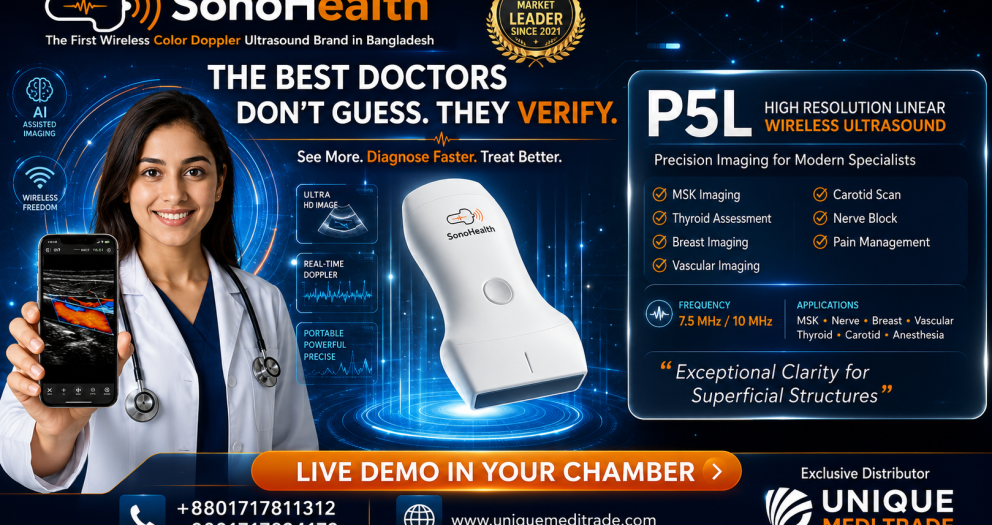
Point-of-Care Ultrasound for Modern Pain Medicine

Nerve Visualization • Musculoskeletal Imaging • Ultrasound-Guided Injection • Needle Guidance • Interventional Pain Procedures

See the Target. Guide the Needle. Treat with Precision.

For physicians, pain clinics and hospitals interested in incorporating Point-of-Care Ultrasound into their Pain Medicine practice: Unique Medi Trade POCUS Solutions • Live Demonstration • Physician Training Support • Technical Support • RFA Pain Management Solutions Contact: +880 1717-811312 Unique Medi Trade — Advancing Image-Guided Pain Medicine in Bangladesh

Medical Disclaimer

This article is intended for healthcare-professional education and general information. Ultrasound-guided pain procedures should only be performed by appropriately trained and credentialed healthcare professionals within their scope of practice and according to relevant clinical guidelines and institutional protocols. Point-of-Care Ultrasound does not replace fluoroscopy, CT, MRI, comprehensive diagnostic ultrasound or specialist imaging when these are clinically indicated. Selection of imaging guidance should depend on the procedure, anatomical target, available evidence, patient condition and physician expertise.    
D2CL SonoHealth Wireless USG

Point-of-Care Ultrasound (POCUS) in Internal Medicine

Point-of-Care Ultrasound (POCUS) in Internal Medicine: Bringing Real-Time Imaging to the Bedside

From “What Do I Think?” to “What Can I See?”

For generations, Internal Medicine physicians have depended on four fundamental tools at the bedside: History → Inspection → Palpation → Auscultation Today, another powerful tool is becoming increasingly relevant: Point-of-Care Ultrasound — POCUS.Point-of-Care Ultrasound (POCUS) in Internal Medicine POCUS enables appropriately trained physicians to perform focused ultrasound examinations at the patient's bedside and integrate real-time imaging findings with the history, physical examination and other clinical information. For an Internal Medicine physician managing patients with shortness of breath, hypotension, edema, acute kidney injury, chest pain, heart failure, pleural effusion or unexplained abdominal distension, immediate ultrasound information can help answer focused clinical questions without always waiting for conventional imaging. The American College of Physicians formally recognizes the important role of POCUS in Internal Medicine and notes its increasing use among internists and subspecialists to improve the timeliness and accuracy of diagnosis. The European Federation of Internal Medicine has similarly described POCUS as a useful tool for internists across emergency departments, hospital wards, consultations and even home-care settings.

What Is Point-of-Care Ultrasound?

Point-of-Care Ultrasound is a focused ultrasound examination performed and interpreted by the treating clinician at or near the point of patient care. The key word is focused. POCUS is usually not intended to reproduce an entire comprehensive echocardiogram, abdominal ultrasound or radiology examination. Instead, it is commonly used to answer specific clinical questions. For example: Does this breathless patient have B-lines? Is there a pleural effusion? Is a pericardial effusion visible? Does gross left ventricular systolic function appear significantly reduced? Is free fluid present in the abdomen? Is urinary retention contributing to acute kidney injury?Point-of-Care Ultrasound (POCUS) in Internal Medicine Is hydronephrosis visible? Could proximal deep-vein thrombosis be present? Where is the safest site for thoracentesis or paracentesis? This question-oriented approach makes POCUS especially relevant to Internal Medicine. A 2025 consensus process involving Internal Medicine POCUS experts identified 12 core diagnostic indications and six procedural indications, with applications including focused cardiac, gallbladder and urinary bladder ultrasound.

Why POCUS Matters in Internal Medicine

Internists frequently manage patients whose symptoms can originate from several organ systems simultaneously. Consider a common presentation:

A 68-year-old patient presents with severe shortness of breath.

The differential diagnosis could include:
  • Acute heart failure
  • Pneumonia
  • Pleural effusion
  • Pneumothorax
  • Pulmonary embolism
  • COPD or asthma exacerbation
  • Pericardial disease
  • Other cardiopulmonary conditions
History, physical examination, ECG, laboratory investigations and formal imaging remain important. But bedside ultrasound may provide additional information within minutes. This ability to integrate ultrasound findings immediately into clinical reasoning is one of the major advantages of POCUS. A 2025 Australian and New Zealand Internal Medicine consensus statement endorsed POCUS in clinical scenarios including fluid-status assessment, undifferentiated shock or hypotension, dyspnea/respiratory failure, acute kidney injury, suspected venous thromboembolism, joint effusion, invasive procedures and guidance of diuretic therapy in heart failure.

1. Lung Ultrasound in Shortness of Breath

Dyspnea is one of the most frequent and challenging presentations in Internal Medicine. A patient with acute breathing difficulty may have a cardiac, pulmonary or systemic cause. With lung POCUS, a trained physician can evaluate findings such as:
  • Lung sliding
  • B-lines
  • Pleural effusion
  • Lung consolidation patterns
  • Findings suggestive of pneumothorax
International evidence-based recommendations on point-of-care lung ultrasound were updated in 2026, reflecting the growing body of evidence and increasingly established role of lung ultrasound in clinical practice.

B-Lines

Multiple B-lines may be seen in conditions associated with increased extravascular lung water, although they are not specific to one disease. For an Internal Medicine physician managing a patient with suspected heart failure, lung ultrasound findings can be interpreted together with: Symptoms + Physical examination + Cardiac POCUS + IVC assessment + Laboratory findings + Other imaging This multi-organ approach can provide a much richer clinical picture than relying on a single finding alone.

2. POCUS in Heart Failure

Heart failure is another major area where POCUS can be useful. Patients may present with:
  • Dyspnea
  • Orthopnea
  • Peripheral edema
  • Fatigue
  • Raised JVP
  • Pulmonary congestion
  • Hypotension
A focused bedside examination may allow the trained internist to assess several complementary areas.

Lung

Are B-lines present? Is there a pleural effusion?Point-of-Care Ultrasound (POCUS) in Internal Medicine

Heart

Does gross left ventricular systolic function appear preserved or significantly reduced? Is pericardial fluid visible? Are there findings that raise concern for right ventricular abnormality?

Inferior Vena Cava

What does the IVC look like in the context of the entire clinical assessment? Used appropriately, these observations can contribute to assessment of congestion and hemodynamic status. The 2025 Internal Medicine Society of Australia and New Zealand consensus specifically supported POCUS for fluid-status assessment and guidance of diuretic therapy in heart failure. Importantly, focused cardiac ultrasound is not equivalent to comprehensive echocardiography. When detailed valve assessment, chamber quantification, Doppler measurements or other advanced evaluation is required, formal echocardiography remains essential.

3. Focused Cardiac Ultrasound

The heart is one of the most important POCUS targets in acute Internal Medicine. Depending on training and clinical context, focused cardiac ultrasound may help answer questions such as:
  • Is there a significant pericardial effusion?
  • Does global LV systolic function appear markedly reduced?Point-of-Care Ultrasound (POCUS) in Internal Medicine
  • Is there obvious right ventricular enlargement?
  • Is the heart hyperdynamic in an appropriate clinical context?
  • Are there findings that may help explain shock or dyspnea?
Internal Medicine POCUS literature includes focused cardiac examination among core domains, and current training consensus frameworks include cardiac ultrasound skills alongside lung, abdominal, procedural and vascular applications. The objective is not: “Perform a complete cardiology echocardiogram.” The objective is: “Answer a focused bedside question that may change immediate clinical management.”

4. POCUS in Undifferentiated Shock and Hypotension

Few clinical situations require faster decision-making than a patient with hypotension or shock. Possible causes may include:
  • HypovolemiaPoint-of-Care Ultrasound (POCUS) in Internal Medicine
  • Sepsis
  • Cardiogenic shock
  • Obstructive processes
  • Hemorrhage
  • Mixed etiologies
POCUS can be particularly powerful in these situations because the physician can evaluate several organ systems rapidly. A focused examination may include:

Heart

Gross ventricular function and pericardial fluid.

Lungs

B-lines, pleural findings or pneumothorax patterns.

IVC

Interpreted carefully within the broader hemodynamic context.

Abdomen

Free fluid where clinically relevant.

Vascular System

Selected venous assessment when thromboembolic disease is suspected. Undifferentiated shock or hypotension is among the clinical scenarios specifically supported in the 2025 Internal Medicine consensus statement. This illustrates one of the greatest strengths of POCUS:

One device can provide focused information from multiple organ systems during the same bedside assessment.


5. Inferior Vena Cava and Volume Assessment

The inferior vena cava, or IVC, is commonly incorporated into bedside ultrasound assessment. An internist may examine:Point-of-Care Ultrasound (POCUS) in Internal Medicine
  • IVC size
  • Respiratory variation
  • The overall clinical context
However, IVC findings should not be interpreted in isolation as a simple measurement of whether a patient is “dry” or “fluid overloaded.” Mechanical ventilation, right-sided cardiac pressures, respiratory effort and several other factors can influence IVC appearance. The best use of POCUS is therefore integrative.

Physical examination

Lung ultrasound

Cardiac findings

IVC findings

Clinical history

Laboratory and other diagnostic information

This combined approach is much more powerful than relying on one ultrasound measurement alone.

6. Pleural Effusion

Pleural effusion is frequently encountered in Internal Medicine. It may occur in association with:
  • Heart failure
  • Infection
  • Malignancy
  • Liver disease
  • Renal disease
  • Other systemic conditions
Bedside ultrasound can help identify pleural fluid and provide information about its location and approximate extent. It can also be used to guide thoracentesis when the procedure is clinically indicated and performed by an appropriately trained physician. The Society of Hospital Medicine has published recommendations related to POCUS for bedside procedures including thoracentesis, paracentesis, vascular access and lumbar puncture. This is an important distinction: POCUS is not only a diagnostic tool. It can also be a procedural guidance tool.

7. Pneumothorax Assessment

Pneumothorax may require rapid recognition, particularly in critically ill or deteriorating patients. Lung ultrasound can evaluate specific sonographic findings associated with pneumothorax. Because the examination can be performed immediately at the bedside and repeated when clinically necessary, POCUS may be particularly useful in acute-care environments. However, sonographic findings must be interpreted by trained operators and integrated with the patient's clinical status and other investigations.

8. Pneumonia and Lung Consolidation

Lung POCUS may also identify peripheral consolidation patterns and associated pleural abnormalities. For an Internal Medicine physician evaluating: Fever + Cough + Dyspnea + Hypoxia POCUS findings can become another component of the diagnostic assessment. It does not eliminate the need for chest radiography, CT, microbiology or other investigations when clinically indicated. Instead, it gives the treating physician another source of real-time bedside information.

9. POCUS in Acute Kidney Injury

Acute kidney injury is a common problem in hospitalized medical patients. The causes may be: Pre-renal → Renal → Post-renal POCUS can contribute particularly to selected questions related to urinary obstruction and bladder volume. A physician may assess:
  • Kidney appearance
  • Possible hydronephrosis
  • Urinary bladder volume
  • Urinary retention
Current Internal Medicine consensus recommendations include acute kidney injury among clinical scenarios where POCUS can provide benefit, with abdominal applications including relevant renal and urinary assessment. Imagine a patient whose creatinine is rapidly rising. Instead of relying only on laboratory values and physical examination, focused bedside imaging may help the clinician determine whether an obvious obstructive process requires further investigation.

10. Bladder Assessment

Portable ultrasound can be particularly practical when urinary retention is suspected. Focused bladder imaging may help estimate whether the bladder is distended and may contribute to decisions about catheterization or further urological evaluation. The 2025 U.S. Internal Medicine residency POCUS consensus specifically included urinary bladder ultrasound and bladder-volume assessment within its recommended applications and skills.

11. Abdominal Free Fluid and Ascites

Patients with chronic liver disease, heart failure, malignancy and other medical conditions may develop ascites. POCUS can help determine:
  • Whether free abdominal fluid is visible
  • Where fluid pockets are located
  • Whether there may be a suitable site for a procedure
For patients requiring paracentesis, ultrasound can also assist in selecting a safer entry site. Earlier Canadian Internal Medicine ultrasound consensus recommendations included abdominal free fluid as a core POCUS application and ultrasound-guided paracentesis among core procedural skills.

12. Gallbladder Assessment

Depending on training and institutional scope, abdominal POCUS may also be used for focused gallbladder assessment. For a patient presenting with: Right upper abdominal pain + Fever + Nausea POCUS may provide additional information regarding gallstones or gallbladder abnormalities. A comprehensive abdominal ultrasound or other diagnostic imaging may still be required depending on the clinical picture. The value of POCUS is that focused information may be available during the initial patient assessment.

13. Deep Vein Thrombosis

A patient presents with: Unilateral leg swelling + Pain + Clinical suspicion of DVT Compression ultrasound performed by an appropriately trained physician can help assess selected proximal veins for suspected deep-vein thrombosis. Evaluation for suspected venous thromboembolism is included among endorsed Internal Medicine POCUS clinical scenarios in contemporary consensus recommendations. A review of Internal Medicine POCUS applications also highlights examination of major venous trunks for proximal venous thrombosis. Depending on the circumstances, comprehensive vascular ultrasound or additional investigation may still be required.

14. Ultrasound-Guided Procedures

One of the most established roles of bedside ultrasound is procedural guidance. Internal Medicine physicians may perform procedures such as:
  • Thoracentesis
  • Paracentesis
  • Central venous access
  • Peripheral vascular access
  • Selected joint procedures
  • Other bedside interventions
Ultrasound allows the physician to visualize anatomy before—or in some cases during—the procedure. For example, before thoracentesis the physician can identify: Chest wall → Pleural space → Fluid → Lung Before paracentesis: Abdominal wall → Peritoneal fluid → Adjacent structures For vascular access: Vein → Artery → Surrounding structures → Needle path The Society of Hospital Medicine emphasizes POCUS for both answering specific diagnostic questions and guiding invasive bedside procedures.

POCUS Changes the Traditional Bedside Workflow

Consider the traditional model: Patient ExaminationUltrasound RequestedPatient Transferred to Imaging DepartmentExamination PerformedReport PreparedPhysician Reviews Result This process remains necessary for many comprehensive diagnostic examinations. But POCUS creates another pathway for appropriately selected clinical questions: Patient ExaminationFocused Bedside UltrasoundImmediate Integration with Clinical FindingsClinical Decision The advantage is not simply speed. It is the ability to correlate imaging with what the physician is observing at that exact moment.

POCUS Is the Visual Extension of the Physical Examination

Consider how medical examination has evolved. A physician can: Listen to the heart with a stethoscope. POCUS may allow the trained physician to look at gross cardiac function. A physician can: Listen to the lungs. POCUS may allow the physician to evaluate for B-lines, pleural fluid or selected lung abnormalities. A physician can: Examine peripheral edema and estimate the JVP. POCUS can provide additional information from the lungs, heart and venous system. A physician can: Palpate the abdomen. POCUS may help visualize ascites, bladder distension, gallbladder findings or selected renal abnormalities. POCUS should therefore not be viewed simply as a smaller version of a radiology ultrasound machine. For the trained internist, it can become a visual extension of bedside clinical examination.

The Power of Multi-Organ POCUS

One of the greatest advantages of POCUS in Internal Medicine is the ability to combine information from multiple organs. For example, consider a patient presenting with acute dyspnea. Instead of asking only: “What do the lungs show?” The physician may evaluate:

Lungs

B-lines? Pleural effusion? Consolidation? Pneumothorax pattern?

Heart

Gross LV function? Pericardial effusion? Right-heart abnormalities?

IVC

What does venous filling look like within the overall context?

Veins

Is DVT assessment indicated? This concept of integrated multi-organ ultrasound fits particularly well with Internal Medicine because internists are trained to treat the patient as a whole rather than focusing exclusively on one organ.

Why Handheld Wireless Ultrasound Is Important for POCUS

POCUS becomes particularly practical when the ultrasound system can move with the physician. Traditional ultrasound machines can provide sophisticated imaging capabilities, but they may be large, expensive and located in designated departments. A handheld wireless ultrasound can make ultrasound accessible in:
  • Medicine wards
  • Doctor's chambers
  • Emergency rooms
  • ICU/HDU
  • Outpatient departments
  • Rural hospitals
  • Mobile medical services
  • Bedside rounds
The physician can potentially carry the device from one patient to another and connect it to a compatible smartphone or tablet. This portability represents a fundamental change in ultrasound workflow.

Ultrasound no longer has to be a place the patient goes.

It can become a tool that comes to the patient.


SonoHealth Handheld Wireless Ultrasound for Internal Medicine POCUS
For Internal Medicine physicians interested in incorporating POCUS into clinical practice, SonoHealth Handheld Wireless Ultrasound offers a portable approach to point-of-care imaging. Depending on the probe configuration, handheld ultrasound can support applications involving:

Convex Probe

Useful for deeper structures and abdominal applications such as:
  • Liver
  • Gallbladder
  • Kidneys
  • Urinary bladder
  • Ascites
  • IVC
  • General abdominal assessment

Phased/Cardiac Probe

Useful for focused cardiac and thoracic applications such as:
  • Focused cardiac assessment
  • Pericardial effusion
  • Gross ventricular function
  • Pleural assessment
  • Lung POCUS

Linear Probe

Useful for superficial and vascular applications including:
  • Vascular access
  • DVT assessment
  • Pleural and superficial structures
  • Soft tissue
  • Selected musculoskeletal applications
A 3-in-1 handheld ultrasound configuration combining Linear + Convex + Cardiac/Phased capabilities can therefore be particularly practical for physicians who want one portable device for multi-organ POCUS.

One Device. Multiple Clinical Questions.

For an Internal Medicine physician, a versatile handheld POCUS system can potentially support focused assessment from: HeartLungIVCAbdomenKidney & BladderPeripheral VeinsProcedural Guidance This is why POCUS is particularly well aligned with Internal Medicine. The specialty itself is multi-system. The ultrasound tool should be equally versatile.

POCUS Does Not Replace Comprehensive Diagnostic Ultrasound

This is an essential principle. Point-of-Care Ultrasound should not be presented as a replacement for radiologists, cardiologists, sonologists or comprehensive diagnostic ultrasound examinations. POCUS generally answers a focused clinical question. A comprehensive examination may require:
  • Detailed imaging protocol
  • Advanced Doppler analysis
  • Multiple standardized measurements
  • Formal echocardiography
  • Detailed organ evaluation
  • Specialist interpretation
  • Structured reporting
  • CT, MRI or other imaging when indicated
Professional ultrasound standards emphasize appropriate indications, operator competence, structured examinations, reporting and quality management. POCUS should complement existing diagnostic pathways—not compete with them.

Training Is More Important Than the Machine

Owning an ultrasound device does not automatically make someone competent in POCUS. Successful implementation requires: Ultrasound Knowledge
Hands-On Training
Supervised Scanning
Image Interpretation
Clinical Integration
Recognition of Limitations Structured training is strongly emphasized by professional organizations. The European Federation of Internal Medicine recommends development of structured POCUS training, while the American College of Physicians provides dedicated foundational and practical POCUS training programs for Internal Medicine physicians. The Society of Hospital Medicine similarly provides POCUS education and competency-focused training pathways for hospitalists. The future of POCUS therefore depends on two things:

Better technology.

and

Better-trained physicians.


POCUS and the Future of Internal Medicine

The stethoscope transformed clinical examination approximately two centuries ago. Today, handheld ultrasound is creating another important evolution in bedside medicine. The goal is not to replace the stethoscope. The goal is not to replace radiology. The goal is not to replace echocardiography. The goal is to give the physician more clinical information at the point of care. International professional organizations are increasingly incorporating POCUS into Internal Medicine practice and education. The ACP formally supports its role in Internal Medicine, the Society of Hospital Medicine has developed position statements and training pathways, and recent consensus recommendations have defined increasingly detailed core applications for Internal Medicine physicians. For the modern internist, the question may therefore be changing from: “Why should I learn POCUS?” to:

“How should I integrate POCUS safely and effectively into my clinical practice?”


See More at the Bedside. Decide with More Information.

Internal Medicine is built around clinical reasoning. POCUS adds real-time visualization to that process. When used by appropriately trained physicians, it can support focused assessment of the: Heart • Lungs • IVC • Abdomen • Kidneys • Bladder • Peripheral Veins and provide ultrasound guidance for selected bedside procedures. The result is not simply a new piece of equipment. It is a different way of approaching bedside medicine.

Listen. Examine. Visualize. Integrate. Decide.


SonoHealth Handheld Wireless Ultrasound

Point-of-Care Ultrasound for Modern Internal Medicine

Portable. Wireless. Multi-Organ Imaging. See More. Diagnose Faster. Treat Better. For physicians, hospitals and institutions interested in introducing or expanding a Point-of-Care Ultrasound program: Unique Medi Trade Clinical Demonstration • POCUS Solutions • Technical Support • Training Support Contact: +880 1717-811312

Medical Disclaimer

This article is intended for healthcare-professional education and general information. Point-of-Care Ultrasound should be performed by appropriately trained clinicians within their scope of practice and according to relevant institutional protocols and professional standards. POCUS findings should always be interpreted together with the patient's clinical history, examination and other investigations. Focused POCUS does not replace comprehensive diagnostic ultrasound, echocardiography, radiology assessment or specialist evaluation when these are clinically indicated.
Back to Top
Product has been added to your cart