Point-of-Care Ultrasound (POCUS) in Internal Medicine: Bringing Real-Time Imaging to the Bedside
From “What Do I Think?” to “What Can I See?”
For generations, Internal Medicine physicians have depended on four fundamental tools at the bedside:
History → Inspection → Palpation → Auscultation
Today, another powerful tool is becoming increasingly relevant:
Point-of-Care Ultrasound — POCUS.
POCUS enables appropriately trained physicians to perform focused ultrasound examinations at the patient's bedside and integrate real-time imaging findings with the history, physical examination and other clinical information.
For an Internal Medicine physician managing patients with
shortness of breath, hypotension, edema, acute kidney injury, chest pain, heart failure, pleural effusion or unexplained abdominal distension, immediate ultrasound information can help answer focused clinical questions without always waiting for conventional imaging.
The American College of Physicians formally recognizes the important role of POCUS in Internal Medicine and notes its increasing use among internists and subspecialists to improve the timeliness and accuracy of diagnosis.
The European Federation of Internal Medicine has similarly described POCUS as a useful tool for internists across emergency departments, hospital wards, consultations and even home-care settings.
What Is Point-of-Care Ultrasound?
Point-of-Care Ultrasound is a
focused ultrasound examination performed and interpreted by the treating clinician at or near the point of patient care.
The key word is
focused.
POCUS is usually not intended to reproduce an entire comprehensive echocardiogram, abdominal ultrasound or radiology examination.
Instead, it is commonly used to answer specific clinical questions.
For example:
Does this breathless patient have B-lines?
Is there a pleural effusion?
Is a pericardial effusion visible?
Does gross left ventricular systolic function appear significantly reduced?
Is free fluid present in the abdomen?
Is urinary retention contributing to acute kidney injury? Is hydronephrosis visible?
Could proximal deep-vein thrombosis be present?
Where is the safest site for thoracentesis or paracentesis?
Is hydronephrosis visible?
Could proximal deep-vein thrombosis be present?
Where is the safest site for thoracentesis or paracentesis?
This question-oriented approach makes POCUS especially relevant to Internal Medicine.
A 2025 consensus process involving Internal Medicine POCUS experts identified
12 core diagnostic indications and six procedural indications, with applications including focused cardiac, gallbladder and urinary bladder ultrasound.
Why POCUS Matters in Internal Medicine
Internists frequently manage patients whose symptoms can originate from several organ systems simultaneously.
Consider a common presentation:
A 68-year-old patient presents with severe shortness of breath.
The differential diagnosis could include:
- Acute heart failure
- Pneumonia
- Pleural effusion
- Pneumothorax
- Pulmonary embolism
- COPD or asthma exacerbation
- Pericardial disease
- Other cardiopulmonary conditions
History, physical examination, ECG, laboratory investigations and formal imaging remain important.
But bedside ultrasound may provide additional information
within minutes.
This ability to integrate ultrasound findings immediately into clinical reasoning is one of the major advantages of POCUS.
A 2025 Australian and New Zealand Internal Medicine consensus statement endorsed POCUS in clinical scenarios including
fluid-status assessment, undifferentiated shock or hypotension, dyspnea/respiratory failure, acute kidney injury, suspected venous thromboembolism, joint effusion, invasive procedures and guidance of diuretic therapy in heart failure.
1. Lung Ultrasound in Shortness of Breath
Dyspnea is one of the most frequent and challenging presentations in Internal Medicine.
A patient with acute breathing difficulty may have a cardiac, pulmonary or systemic cause.
With lung POCUS, a trained physician can evaluate findings such as:
- Lung sliding
- B-lines
- Pleural effusion
- Lung consolidation patterns
- Findings suggestive of pneumothorax
International evidence-based recommendations on point-of-care lung ultrasound were updated in 2026, reflecting the growing body of evidence and increasingly established role of lung ultrasound in clinical practice.
B-Lines
Multiple B-lines may be seen in conditions associated with increased extravascular lung water, although they are not specific to one disease.
For an Internal Medicine physician managing a patient with suspected heart failure, lung ultrasound findings can be interpreted together with:
Symptoms + Physical examination + Cardiac POCUS + IVC assessment + Laboratory findings + Other imaging
This multi-organ approach can provide a much richer clinical picture than relying on a single finding alone.
2. POCUS in Heart Failure
Heart failure is another major area where POCUS can be useful.
Patients may present with:
- Dyspnea
- Orthopnea
- Peripheral edema
- Fatigue
- Raised JVP
- Pulmonary congestion
- Hypotension
A focused bedside examination may allow the trained internist to assess several complementary areas.
Lung
Are B-lines present?
Is there a pleural effusion?

Heart
Does gross left ventricular systolic function appear preserved or significantly reduced?
Is pericardial fluid visible?
Are there findings that raise concern for right ventricular abnormality?
Inferior Vena Cava
What does the IVC look like in the context of the entire clinical assessment?
Used appropriately, these observations can contribute to assessment of congestion and hemodynamic status.
The 2025 Internal Medicine Society of Australia and New Zealand consensus specifically supported POCUS for
fluid-status assessment and guidance of diuretic therapy in heart failure.
Importantly, focused cardiac ultrasound is not equivalent to comprehensive echocardiography.
When detailed valve assessment, chamber quantification, Doppler measurements or other advanced evaluation is required, formal echocardiography remains essential.
3. Focused Cardiac Ultrasound
The heart is one of the most important POCUS targets in acute Internal Medicine.
Depending on training and clinical context, focused cardiac ultrasound may help answer questions such as:
- Is there a significant pericardial effusion?
- Does global LV systolic function appear markedly reduced?

- Is there obvious right ventricular enlargement?
- Is the heart hyperdynamic in an appropriate clinical context?
- Are there findings that may help explain shock or dyspnea?
Internal Medicine POCUS literature includes focused cardiac examination among core domains, and current training consensus frameworks include cardiac ultrasound skills alongside lung, abdominal, procedural and vascular applications.
The objective is not:
“Perform a complete cardiology echocardiogram.”
The objective is:
“Answer a focused bedside question that may change immediate clinical management.”
4. POCUS in Undifferentiated Shock and Hypotension
Few clinical situations require faster decision-making than a patient with hypotension or shock.
Possible causes may include:
- Hypovolemia

- Sepsis
- Cardiogenic shock
- Obstructive processes
- Hemorrhage
- Mixed etiologies
POCUS can be particularly powerful in these situations because the physician can evaluate several organ systems rapidly.
A focused examination may include:
Heart
Gross ventricular function and pericardial fluid.
Lungs
B-lines, pleural findings or pneumothorax patterns.
IVC
Interpreted carefully within the broader hemodynamic context.
Abdomen
Free fluid where clinically relevant.
Vascular System
Selected venous assessment when thromboembolic disease is suspected.
Undifferentiated shock or hypotension is among the clinical scenarios specifically supported in the 2025 Internal Medicine consensus statement.
This illustrates one of the greatest strengths of POCUS:
One device can provide focused information from multiple organ systems during the same bedside assessment.
5. Inferior Vena Cava and Volume Assessment
The inferior vena cava, or IVC, is commonly incorporated into bedside ultrasound assessment.
An internist may examine:
- IVC size
- Respiratory variation
- The overall clinical context
However, IVC findings should
not be interpreted in isolation as a simple measurement of whether a patient is “dry” or “fluid overloaded.”
Mechanical ventilation, right-sided cardiac pressures, respiratory effort and several other factors can influence IVC appearance.
The best use of POCUS is therefore integrative.
Physical examination
Lung ultrasound
Cardiac findings
IVC findings
Clinical history
Laboratory and other diagnostic information
This combined approach is much more powerful than relying on one ultrasound measurement alone.
6. Pleural Effusion
Pleural effusion is frequently encountered in Internal Medicine.
It may occur in association with:
- Heart failure
- Infection
- Malignancy
- Liver disease
- Renal disease
- Other systemic conditions
Bedside ultrasound can help identify pleural fluid and provide information about its location and approximate extent.
It can also be used to guide thoracentesis when the procedure is clinically indicated and performed by an appropriately trained physician.
The Society of Hospital Medicine has published recommendations related to POCUS for bedside procedures including
thoracentesis, paracentesis, vascular access and lumbar puncture.
This is an important distinction:
POCUS is not only a
diagnostic tool.
It can also be a
procedural guidance tool.
7. Pneumothorax Assessment
Pneumothorax may require rapid recognition, particularly in critically ill or deteriorating patients.
Lung ultrasound can evaluate specific sonographic findings associated with pneumothorax.
Because the examination can be performed immediately at the bedside and repeated when clinically necessary, POCUS may be particularly useful in acute-care environments.
However, sonographic findings must be interpreted by trained operators and integrated with the patient's clinical status and other investigations.
8. Pneumonia and Lung Consolidation
Lung POCUS may also identify peripheral consolidation patterns and associated pleural abnormalities.
For an Internal Medicine physician evaluating:
Fever + Cough + Dyspnea + Hypoxia
POCUS findings can become another component of the diagnostic assessment.
It does not eliminate the need for chest radiography, CT, microbiology or other investigations when clinically indicated.
Instead, it gives the treating physician another source of
real-time bedside information.
9. POCUS in Acute Kidney Injury
Acute kidney injury is a common problem in hospitalized medical patients.
The causes may be:
Pre-renal → Renal → Post-renal
POCUS can contribute particularly to selected questions related to urinary obstruction and bladder volume.
A physician may assess:
- Kidney appearance
- Possible hydronephrosis
- Urinary bladder volume
- Urinary retention
Current Internal Medicine consensus recommendations include acute kidney injury among clinical scenarios where POCUS can provide benefit, with abdominal applications including relevant renal and urinary assessment.
Imagine a patient whose creatinine is rapidly rising.
Instead of relying only on laboratory values and physical examination, focused bedside imaging may help the clinician determine whether an obvious obstructive process requires further investigation.
10. Bladder Assessment
Portable ultrasound can be particularly practical when urinary retention is suspected.
Focused bladder imaging may help estimate whether the bladder is distended and may contribute to decisions about catheterization or further urological evaluation.
The 2025 U.S. Internal Medicine residency POCUS consensus specifically included
urinary bladder ultrasound and bladder-volume assessment within its recommended applications and skills.
11. Abdominal Free Fluid and Ascites
Patients with chronic liver disease, heart failure, malignancy and other medical conditions may develop ascites.
POCUS can help determine:
- Whether free abdominal fluid is visible
- Where fluid pockets are located
- Whether there may be a suitable site for a procedure
For patients requiring paracentesis, ultrasound can also assist in selecting a safer entry site.
Earlier Canadian Internal Medicine ultrasound consensus recommendations included
abdominal free fluid as a core POCUS application and ultrasound-guided
paracentesis among core procedural skills.
12. Gallbladder Assessment
Depending on training and institutional scope, abdominal POCUS may also be used for focused gallbladder assessment.
For a patient presenting with:
Right upper abdominal pain + Fever + Nausea
POCUS may provide additional information regarding gallstones or gallbladder abnormalities.
A comprehensive abdominal ultrasound or other diagnostic imaging may still be required depending on the clinical picture.
The value of POCUS is that focused information may be available
during the initial patient assessment.
13. Deep Vein Thrombosis
A patient presents with:
Unilateral leg swelling + Pain + Clinical suspicion of DVT
Compression ultrasound performed by an appropriately trained physician can help assess selected proximal veins for suspected deep-vein thrombosis.
Evaluation for suspected venous thromboembolism is included among endorsed Internal Medicine POCUS clinical scenarios in contemporary consensus recommendations.
A review of Internal Medicine POCUS applications also highlights examination of major venous trunks for proximal venous thrombosis.
Depending on the circumstances, comprehensive vascular ultrasound or additional investigation may still be required.
14. Ultrasound-Guided Procedures
One of the most established roles of bedside ultrasound is procedural guidance.
Internal Medicine physicians may perform procedures such as:
- Thoracentesis
- Paracentesis
- Central venous access
- Peripheral vascular access
- Selected joint procedures
- Other bedside interventions
Ultrasound allows the physician to visualize anatomy before—or in some cases during—the procedure.
For example, before thoracentesis the physician can identify:
Chest wall → Pleural space → Fluid → Lung
Before paracentesis:
Abdominal wall → Peritoneal fluid → Adjacent structures
For vascular access:
Vein → Artery → Surrounding structures → Needle path
The Society of Hospital Medicine emphasizes POCUS for both answering specific diagnostic questions and guiding invasive bedside procedures.
POCUS Changes the Traditional Bedside Workflow
Consider the traditional model:
Patient Examination
↓
Ultrasound Requested
↓
Patient Transferred to Imaging Department
↓
Examination Performed
↓
Report Prepared
↓
Physician Reviews Result
This process remains necessary for many comprehensive diagnostic examinations.
But POCUS creates another pathway for appropriately selected clinical questions:
Patient Examination
↓
Focused Bedside Ultrasound
↓
Immediate Integration with Clinical Findings
↓
Clinical Decision
The advantage is not simply speed.
It is the ability to correlate imaging with what the physician is observing
at that exact moment.
POCUS Is the Visual Extension of the Physical Examination
Consider how medical examination has evolved.
A physician can:
Listen to the heart with a stethoscope.
POCUS may allow the trained physician to
look at gross cardiac function.
A physician can:
Listen to the lungs.
POCUS may allow the physician to evaluate for
B-lines, pleural fluid or selected lung abnormalities.
A physician can:
Examine peripheral edema and estimate the JVP.
POCUS can provide additional information from the
lungs, heart and venous system.
A physician can:
Palpate the abdomen.
POCUS may help visualize
ascites, bladder distension, gallbladder findings or selected renal abnormalities.
POCUS should therefore not be viewed simply as a smaller version of a radiology ultrasound machine.
For the trained internist, it can become a
visual extension of bedside clinical examination.
The Power of Multi-Organ POCUS
One of the greatest advantages of POCUS in Internal Medicine is the ability to combine information from multiple organs.
For example, consider a patient presenting with acute dyspnea.
Instead of asking only:
“What do the lungs show?”
The physician may evaluate:
Lungs
B-lines? Pleural effusion? Consolidation? Pneumothorax pattern?
Heart
Gross LV function? Pericardial effusion? Right-heart abnormalities?
IVC
What does venous filling look like within the overall context?
Veins
Is DVT assessment indicated?
This concept of
integrated multi-organ ultrasound fits particularly well with Internal Medicine because internists are trained to treat the patient as a whole rather than focusing exclusively on one organ.
Why Handheld Wireless Ultrasound Is Important for POCUS
POCUS becomes particularly practical when the ultrasound system can move with the physician.
Traditional ultrasound machines can provide sophisticated imaging capabilities, but they may be large, expensive and located in designated departments.
A handheld wireless ultrasound can make ultrasound accessible in:
- Medicine wards
- Doctor's chambers
- Emergency rooms
- ICU/HDU
- Outpatient departments
- Rural hospitals
- Mobile medical services
- Bedside rounds
The physician can potentially carry the device from one patient to another and connect it to a compatible smartphone or tablet.
This portability represents a fundamental change in ultrasound workflow.
Ultrasound no longer has to be a place the patient goes.
It can become a tool that comes to the patient.
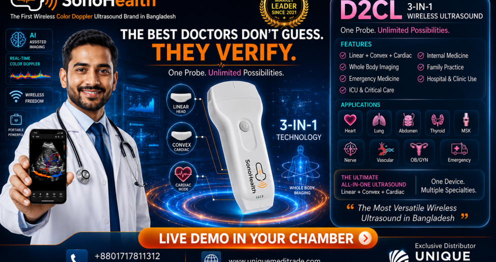
SonoHealth Handheld Wireless Ultrasound for Internal Medicine POCUS
For Internal Medicine physicians interested in incorporating POCUS into clinical practice,
SonoHealth Handheld Wireless Ultrasound offers a portable approach to point-of-care imaging.
Depending on the probe configuration, handheld ultrasound can support applications involving:
Convex Probe
Useful for deeper structures and abdominal applications such as:
- Liver
- Gallbladder
- Kidneys
- Urinary bladder
- Ascites
- IVC
- General abdominal assessment
Phased/Cardiac Probe
Useful for focused cardiac and thoracic applications such as:
- Focused cardiac assessment
- Pericardial effusion
- Gross ventricular function
- Pleural assessment
- Lung POCUS
Linear Probe
Useful for superficial and vascular applications including:
- Vascular access
- DVT assessment
- Pleural and superficial structures
- Soft tissue
- Selected musculoskeletal applications
A
3-in-1 handheld ultrasound configuration combining Linear + Convex + Cardiac/Phased capabilities can therefore be particularly practical for physicians who want one portable device for multi-organ POCUS.
One Device. Multiple Clinical Questions.
For an Internal Medicine physician, a versatile handheld POCUS system can potentially support focused assessment from:
Heart
↓
Lung
↓
IVC
↓
Abdomen
↓
Kidney & Bladder
↓
Peripheral Veins
↓
Procedural Guidance
This is why POCUS is particularly well aligned with Internal Medicine.
The specialty itself is multi-system.
The ultrasound tool should be equally versatile.
POCUS Does Not Replace Comprehensive Diagnostic Ultrasound
This is an essential principle.
Point-of-Care Ultrasound should
not be presented as a replacement for radiologists, cardiologists, sonologists or comprehensive diagnostic ultrasound examinations.
POCUS generally answers a focused clinical question.
A comprehensive examination may require:
- Detailed imaging protocol
- Advanced Doppler analysis
- Multiple standardized measurements
- Formal echocardiography
- Detailed organ evaluation
- Specialist interpretation
- Structured reporting
- CT, MRI or other imaging when indicated
Professional ultrasound standards emphasize appropriate indications, operator competence, structured examinations, reporting and quality management.
POCUS should complement existing diagnostic pathways—not compete with them.
Training Is More Important Than the Machine
Owning an ultrasound device does not automatically make someone competent in POCUS.
Successful implementation requires:
Ultrasound Knowledge
Hands-On Training
Supervised Scanning
Image Interpretation
Clinical Integration
Recognition of Limitations
Structured training is strongly emphasized by professional organizations.
The European Federation of Internal Medicine recommends development of structured POCUS training, while the American College of Physicians provides dedicated foundational and practical POCUS training programs for Internal Medicine physicians.
The Society of Hospital Medicine similarly provides POCUS education and competency-focused training pathways for hospitalists.
The future of POCUS therefore depends on two things:
Better technology.
and
Better-trained physicians.
POCUS and the Future of Internal Medicine
The stethoscope transformed clinical examination approximately two centuries ago.
Today, handheld ultrasound is creating another important evolution in bedside medicine.
The goal is not to replace the stethoscope.
The goal is not to replace radiology.
The goal is not to replace echocardiography.
The goal is to give the physician
more clinical information at the point of care.
International professional organizations are increasingly incorporating POCUS into Internal Medicine practice and education. The ACP formally supports its role in Internal Medicine, the Society of Hospital Medicine has developed position statements and training pathways, and recent consensus recommendations have defined increasingly detailed core applications for Internal Medicine physicians.
For the modern internist, the question may therefore be changing from:
“Why should I learn POCUS?”
to:
“How should I integrate POCUS safely and effectively into my clinical practice?”
See More at the Bedside. Decide with More Information.
Internal Medicine is built around clinical reasoning.
POCUS adds real-time visualization to that process.
When used by appropriately trained physicians, it can support focused assessment of the:
Heart • Lungs • IVC • Abdomen • Kidneys • Bladder • Peripheral Veins
and provide ultrasound guidance for selected bedside procedures.
The result is not simply a new piece of equipment.
It is a different way of approaching bedside medicine.
Listen. Examine. Visualize. Integrate. Decide.
SonoHealth Handheld Wireless Ultrasound
Point-of-Care Ultrasound for Modern Internal Medicine
Portable. Wireless. Multi-Organ Imaging.
See More. Diagnose Faster. Treat Better.
For physicians, hospitals and institutions interested in introducing or expanding a Point-of-Care Ultrasound program:
Unique Medi Trade
Clinical Demonstration • POCUS Solutions • Technical Support • Training Support
Contact: +880 1717-811312
Medical Disclaimer
This article is intended for healthcare-professional education and general information. Point-of-Care Ultrasound should be performed by appropriately trained clinicians within their scope of practice and according to relevant institutional protocols and professional standards. POCUS findings should always be interpreted together with the patient's clinical history, examination and other investigations. Focused POCUS does not replace comprehensive diagnostic ultrasound, echocardiography, radiology assessment or specialist evaluation when these are clinically indicated.