Search

Handheld Portable USG

Portable Ultrasound Machine: Key Considerations for Doctor

Here are 30 key considerations for choosing a portable ultrasound machine for doctors, covering aspects from clinical requirements to budget and technical specifications:

Clinical Requirements

  1. Intended Use: Determine the primary applications (e.g., OB/GYN, cardiac, MSK, emergency) as each requires specific imaging capabilities.
  2. Imaging Modes: Check for the availability of necessary imaging modes (B-mode, M-mode, color Doppler, etc.).
  3. Patient Volume: Consider if the device will handle high patient volumes, as more durable, faster devices are needed for busy practices.
  4. Workflow Efficiency: Opt for models that allow easy patient data entry, annotation, and quick access to settings.
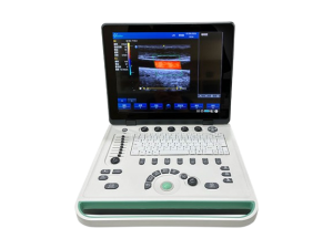
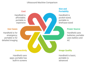
  5. Portability Needs: Choose between handheld, laptop-sized, or trolley-mounted based on how frequently the device will be moved.

Double Headed

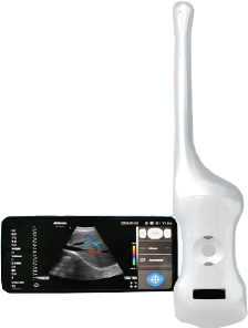
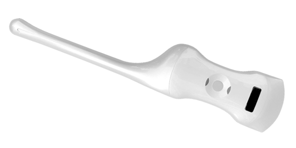
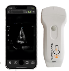
Double Headed Linear + Convex Handheld Wireless Color Doppler Ultrasound​

Technical Specifications

  1. Image Quality: High-resolution imaging is essential for accurate diagnosis, especially for fine details in MSK, vascular, or cardiac scans.
  2. Frequency Range: Consider probes with adjustable frequencies to adapt to different tissue depths (e.g., superficial structures vs. abdominal organs).
  3. Doppler Capabilities: Color, power, and spectral Doppler options are crucial for vascular, cardiac, and obstetric assessments.
  4. Probe Types and Versatility: Ensure the machine has compatible probes for your specialty, such as linear, convex, and phased array probes.
  5. Battery Life: Look for models with long battery life for uninterrupted scanning, particularly for field or emergency use.

Connectivity and Integration

  1. Wireless Capability: Wireless options are convenient for mobile use and remote diagnostics, ideal for connecting with smartphones or tablets.
  2. Data Storage and Transfer: Evaluate storage capacity and transfer options like cloud integration, USB, Wi-Fi, or DICOM for record-keeping.
  3. Compatibility with Devices: If using a handheld device, ensure compatibility with your preferred mobile devices (iOS or Android).
  4. Software Updates: Check if the machine supports software updates to keep it up-to-date with the latest imaging technology.
  5. Connectivity with EMR Systems: Seamless integration with Electronic Medical Records (EMR) systems is crucial for efficient patient data management.
Handheld Portable USG

Handheld Portable USG

User Interface and Ease of Use

  1. User-Friendly Interface: Touchscreen and intuitive menu options improve ease of use, particularly for busy clinicians.
  2. Preset Configurations: Machines with preset imaging settings can save time and enhance consistency in common exams.
  3. Touch and Voice Control: Some portable models offer touch or voice control for hands-free operation.
  4. Quick Boot-Up Time: Short boot-up time is essential for emergency and point-of-care applications where time is critical.
  5. Customizable Settings: Ability to adjust gain, depth, and contrast for various clinical applications enhances versatility.

Physical Design and Durability

  1. Weight and Size: Lighter models are easier to carry and operate for doctors who perform mobile or remote diagnostics.
  2. Build Quality: Durable materials and rugged designs are critical for use in challenging environments.
  3. Waterproof/Resistant Options: Waterproof models are valuable in emergency settings and for hygiene maintenance.
  4. Drop Resistance: Some handheld devices are drop-resistant, which is useful for mobile clinicians.
  5. Ergonomic Design: Comfortable design reduces strain during prolonged use, especially for handheld devices.

Additional Features and Support

  1. Artificial Intelligence (AI) Assistance: AI-based image interpretation or assistance can enhance diagnostic accuracy and speed.
  2. Manufacturer Support and Warranty: Look for reliable technical support and a warranty that covers essential repairs.
  3. Cost of Ownership: Factor in the long-term costs of consumables, maintenance, and software upgrades.
  4. Training and User Resources: Check if the manufacturer offers training or resources, as some devices have a learning curve.
  5. Budget and Financing Options: Balance your budget with features; some suppliers offer financing plans for high-quality machines.

Bonus Tips

  • Feedback from Colleagues: Hearing from other clinicians who use the device can provide practical insights.
  • Demonstration Options: Many suppliers offer demo units or trials, which can be invaluable for evaluating comfort and performance.
Carefully evaluating these considerations can help doctors choose the best portable ultrasound machine for their clinical, financial, and operational needs.

Wireless Portable Ultrasound in Physical Medicine

Wireless Portable Ultrasound in Physical Medicine: Transforming Injury Diagnosis and Treatment on the Go

In physical medicine, wireless portable ultrasounds are highly beneficial for musculoskeletal (MSK) diagnostics, injury management, and procedural guidance. Here’s how they’re used across different applications:

Key Applications of Wireless Portable Ultrasound in Physical Medicine

  1. Musculoskeletal Injury Assessment: Identifies soft tissue injuries, including muscle tears, ligament sprains, tendon injuries, and joint abnormalities. Portable ultrasound allows for real-time imaging to assess the extent of the injury, which aids in developing a treatment plan.
  2. Dynamic Imaging for Functional Assessments: Captures images of joints, tendons, and muscles during movement, helping assess functional impairments and providing valuable insight into movement-related issues like rotator cuff injuries or knee instability.
  3. Guided Injections and Interventions: Ensures precision when performing joint injections, aspiration, or tendon sheath injections. Ultrasound guidance reduces the risk of complications, provides pain relief more effectively, and allows for accurate targeting of the affected area.
  4. Monitoring Healing and Rehabilitation: Tracks tissue recovery over time, allowing for the adjustment of rehabilitation programs based on the healing progress. It’s valuable for conditions like tendinopathies or chronic muscle injuries, where ongoing evaluation is necessary.
  5. Joint Effusion and Inflammation Detection: Visualizes fluid buildup within joints, helping identify conditions like synovitis, bursitis, and arthritis, which are common in patients with chronic joint pain.
  6. Nerve Imaging for Pain Management: Helps locate nerves and assess any structural abnormalities, guiding interventions such as nerve blocks or dry needling for pain management in conditions like carpal tunnel syndrome or sciatic pain.
  7. Evaluation of Chronic Conditions: Provides ongoing assessment for chronic conditions like rheumatoid arthritis or osteoarthritis, helping track changes in joint and soft tissue over time and guiding treatment adjustments.
  8. Pre- and Post-Procedure Evaluation: Used before and after procedures to ensure that treatment targets were achieved and to monitor outcomes, essential for interventions such as platelet-rich plasma (PRP) injections or regenerative medicine procedures.
  9. Educational Tool for Patients: Allows patients to view real-time images of their injuries, improving understanding of their condition and compliance with treatment plans. This visual feedback can increase patient engagement in their recovery.
  10. Portable, On-Site Assessments in Various Settings: Enables MSK assessments in various environments such as sports fields, rehabilitation centers, and even home visits, allowing for convenient, immediate diagnostics and care.

Advantages of Wireless Portable Ultrasound in Physical Medicine

  • Mobility: Wireless technology offers the flexibility to assess patients in various settings, making it ideal for mobile clinics, sports events, and outreach programs.
  • Enhanced Patient Comfort: Real-time imaging minimizes discomfort as patients can undergo immediate, non-invasive assessment and see treatment progress directly.
  • Cost-Effectiveness: Portable ultrasound is a more affordable imaging option than MRI or CT, making it accessible for frequent assessments and follow-ups.
Overall, wireless portable ultrasound is a game-changer in physical medicine, offering precise diagnostics, enhancing patient care, and supporting rehabilitation with real-time, on-the-go imaging.
Handheld Wireless Portable Color Doppler Ultrasound

Wireless Portable Ultrasounds for Emergency Medicine

Wireless portable ultrasounds are used in emergency medicine for a variety of applications, including:
1. Trauma diagnosis: Portable ultrasounds can be used to diagnose abdominal bleeding, limb fractures, and other trauma conditions. They can also be used to guide emergency operations like tracheal intubation and pericardial cavity puncture.
2. Critical condition assessment: Portable ultrasounds can quickly assess for critical conditions like pneumothorax, pericardial effusion, and abdominal bleeding.
3. Cardiac arrest monitoring: Portable ultrasounds can be used to monitor patient changes during cardiac arrest.
4. Deep vein thrombosis evaluation: Portable ultrasounds can be used to rapidly evaluate patients for deep vein thrombosis.
5. Procedural safety enhancement: Portable ultrasounds can be used to enhance procedural safety.
6. Soft tissue and bone damage detection: Portable ultrasounds can detect the degree of soft tissue and bone damage at a trauma site.
7. Fluid accumulation assessment: Portable ultrasounds can assess fluid accumulation in joint cavities.
8. Abnormal muscle echogenicity assessment: Portable ultrasounds can assess abnormal muscle echogenicity.
9. Pneumothorax Detection: Helps detect collapsed lungs in trauma cases or patients with respiratory distress, enabling fast intervention.
10. Vascular Access: Provides guidance for the accurate placement of central or peripheral intravenous lines in difficult-to-access patients, ensuring safer and quicker access.
11. Pulmonary Assessment: Detects pleural effusion (fluid in the lungs), pulmonary edema, or pneumonia, supporting respiratory management decisions.
12. Ectopic Pregnancy Detection: Identifies ectopic pregnancies in women presenting with abdominal pain or shock, allowing for timely surgical intervention.
13. Guidance for Emergency Procedures: Assists with needle placement for procedures like thoracentesis (removing fluid from the lungs) or pericardiocentesis (removing fluid from around the heart).
14 .Fracture Assessment: Visualizes bone fractures, especially in remote or pre-hospital settings where X-ray access may be limited.
These applications make wireless portable ultrasound a critical tool in emergency medicine, enabling rapid, non-invasive diagnostics that can guide timely and potentially life-saving interventions.
Contact: +8801717811312

Wireless Portable Ultrasound is Very Essential physician Tools for Emergency Situation

Wireless portable ultrasound has indeed become an essential tool for physicians in emergency situations, offering quick, accurate, and non-invasive diagnostic capabilities. Its significance is clear across multiple emergency scenarios:

Key Reasons Wireless Portable Ultrasound is Essential in Emergencies

  1. Rapid Diagnosis at the Point of Care: Wireless ultrasound allows physicians to quickly assess critical conditions like internal bleeding, cardiac arrest, pneumothorax (collapsed lung), and abdominal injuries directly at the bedside, in ambulances, or even in remote settings.
  2. Mobility and Convenience: Wireless devices are compact and easy to carry, allowing physicians to perform diagnostics wherever the patient is located. This mobility is crucial for emergency responders in ambulances, paramedics on-site, or military medics in field hospitals.
  3. No Need for External Power or Cables: Wireless devices typically operate on rechargeable batteries and don’t rely on wall outlets or cumbersome cables, providing flexibility in fast-paced, chaotic environments.
  4. Real-Time Imaging for Life-Saving Interventions: Wireless ultrasound offers live imaging for interventions like central line placement, needle thoracostomy for collapsed lungs, and guiding resuscitation efforts in cardiac arrest. This accuracy can significantly improve patient outcomes in time-sensitive scenarios.
  5. Better Patient Outcomes Through Immediate Decision-Making: Physicians can make quick decisions on patient stabilization, surgical need, or transfer based on ultrasound findings. The device also helps to differentiate between similar symptoms (e.g., chest pain due to heart vs. lung issues), directing treatment more accurately.
  6. Data Sharing and Remote Collaboration: Wireless ultrasound devices often connect to mobile devices or hospital networks, allowing images and findings to be shared instantly with specialists, even if they’re off-site. This capability improves diagnostic accuracy and supports collaborative decision-making, which is vital in complex emergencies.

Common Emergency Applications for Wireless Portable Ultrasound

  • Trauma Assessment (FAST exam): Evaluating abdominal and chest trauma for internal bleeding.
  • Cardiac Emergencies: Assessing heart function and detecting pericardial effusion (fluid around the heart).
  • Lung Evaluation: Detecting pneumothorax or pleural effusion (fluid in the lungs).
  • Abdominal Pain: Quick assessment for appendicitis, gallstones, or ectopic pregnancy.
  • Vascular Access: Assisting with intravenous and central line placements.
Overall, wireless portable ultrasound has become indispensable in emergency medicine, offering the speed, accuracy, and flexibility that are crucial in saving lives during critical moments.
Back to Top
Product has been added to your cart