Search

Application & Benefits of Endorectal Handheld Wireless Color Doppler Ultrasound

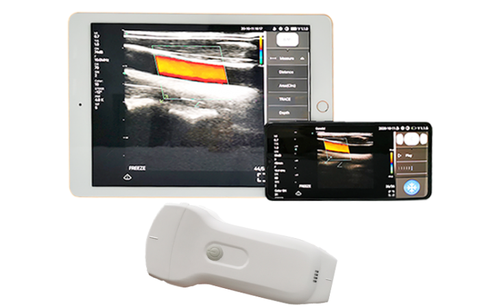
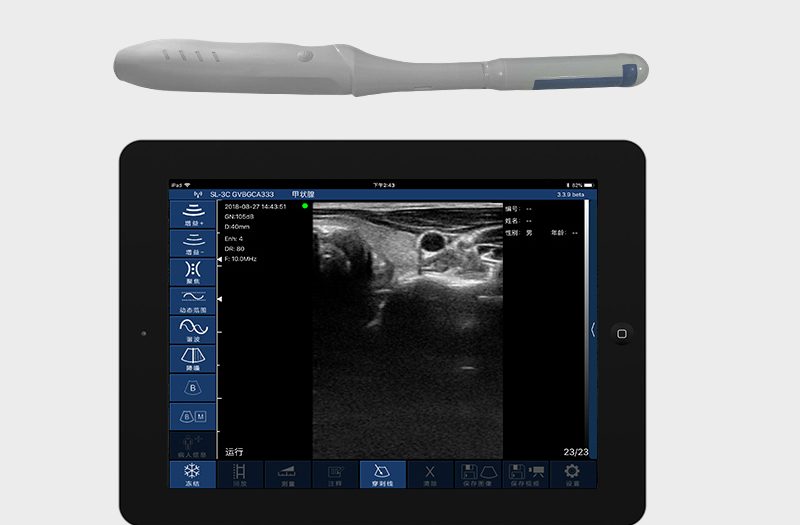
The endorectal handheld wireless color Doppler ultrasound is a versatile and portable diagnostic tool with many applications and benefits in modern medical practice. Below are detailed applications and benefits of this device:

Contact for details:

WhatsApp/Call: 01717811312


Applications

1. Prostate Imaging

  • Diagnosis: Identifying prostate enlargement (BPH) or cancer.
  • Monitoring: Assessing changes in prostate size or vascularity over time.

2. Rectal Cancer Diagnosis and Staging

  • Determining tumor size, invasion depth, and involvement of lymph nodes or surrounding tissues.

3. Evaluation of Pelvic Floor Dysfunction

  • Assessing rectal prolapse, fecal incontinence, or muscle weakening.

4. Inflammatory Bowel Disease (IBD)

  • Visualizing rectal involvement in Crohn’s disease or ulcerative colitis.

5. Anal Fistula and Abscess Imaging

  • Mapping fistula tracts and identifying abscesses to aid surgical planning.

6. Post-Surgical Monitoring

  • Ensuring proper healing and detecting complications after rectal or prostate surgeries.

7. Biopsy Guidance

  • Assisting in precise needle placement for prostate or rectal biopsies.

8. Radiotherapy Planning

  • Providing detailed imaging to guide radiation treatment for rectal or prostate cancer.

9. Sexual Dysfunction Evaluation

  • Assessing vascular abnormalities and pelvic blood flow related to erectile dysfunction.

10. Pelvic Tumor Assessment

  • Identifying benign or malignant tumors in the rectal or pelvic regions.

11. Hemorrhoids and Vascular Disorders

  • Evaluating blood flow in hemorrhoids and rectal varices using Doppler imaging.

12. Rectal Foreign Body Detection

  • Localizing foreign objects in the rectum for safe and effective removal.

13. Ischemic Bowel Diagnosis

  • Evaluating blood flow to detect ischemia in the rectal area.

14. Endometriosis

  • Assessing rectal wall involvement in pelvic endometriosis.

15. Pediatric Anomalies

  • Visualizing congenital malformations of the rectum or pelvic region.

16. Rectocele Assessment

  • Diagnosing rectocele in women with pelvic organ prolapse.

17. Post-Radiation Monitoring

  • Detecting radiation-induced tissue damage or cancer recurrence.

18. Chronic Anorectal Pain

  • Identifying structural or vascular causes of persistent rectal pain.

19. Assisted Therapies

  • Real-time imaging during treatments like prostate brachytherapy or abscess drainage.

20. Training and Research

  • Used in medical training for understanding rectal and pelvic anatomy.

Benefits

1. Portability

  • Compact and lightweight, making it ideal for bedside, outpatient clinics, or remote locations.

2. Wireless Connectivity

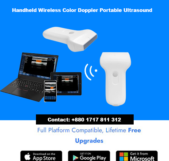
  • Real-time image transfer to smartphones, tablets, or cloud storage for remote interpretation or sharing with specialists.

3. Cost-Effectiveness

  • More affordable than traditional cart-based ultrasound machines, lowering diagnostic costs for patients and clinics.

4. High-Quality Imaging

  • Provides clear 2D, and Color Doppler images, crucial for evaluating blood flow and detecting abnormalities.

5. Non-Invasive

  • Offers a safe and minimally invasive way to evaluate rectal and pelvic organs.

6. Real-Time Imaging

  • Enables immediate diagnosis and dynamic evaluation of blood flow and organ movement.

7. Ease of Use

  • User-friendly design with intuitive controls, reducing the learning curve for healthcare professionals.

8. Improved Patient Comfort

  • Compact probe size minimizes patient discomfort during rectal exams compared to larger devices.

9. Versatility

  • Can be used across a wide range of clinical settings, from general practice to specialized oncology and gastroenterology clinics.

10. Enhanced Workflow

  • Quick setup and wireless functionality improve efficiency, allowing clinicians to perform more procedures in less time.
Portable Ultrasound

Basic difference between handheld ultrasound machines (Handheld USG) and portable ultrasound machines

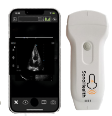
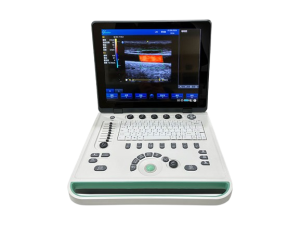
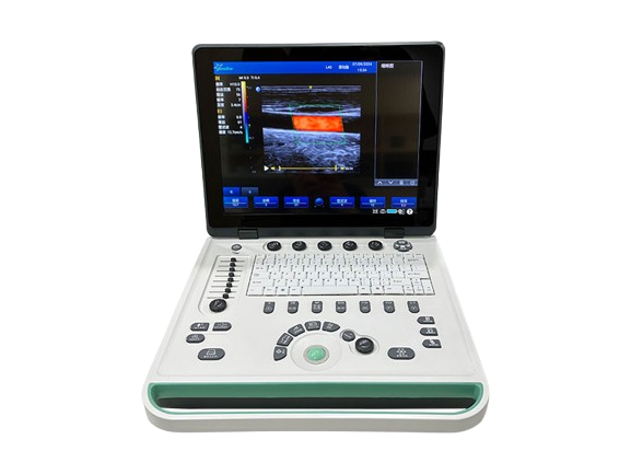
The basic difference between handheld ultrasound machines (Handheld USG) and portable ultrasound machines lies in their size, functionality, and use cases. Here's a detailed comparison: Handheld Wireless Portable Color Doppler UltrasoundPortable Ultrasound  

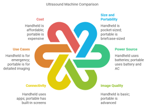
1. Size and Portability

  • Handheld USG:
    • Extremely compact and lightweight, often resembling a smartphone or a small tablet.
    • Fits into a pocket, making it easy to carry and use on the go.
  • Portable USG:
    • Larger than handheld devices, typically like a laptop or a small briefcase.
    • Portable but requires a dedicated bag or trolley for easier mobility.

2. Power Source

  • Handheld USG:
    • Usually powered by rechargeable batteries, offering a few hours of operation.
    • Completely wireless, with data transferred via Wi-Fi or Bluetooth.
  • Portable USG:
    • Can operate on both battery and AC power.
    • Often comes with built-in power systems for longer usage times in clinics or hospitals.

3. Image Quality

  • Handheld USG:
    • Advanced handheld devices, like color Doppler models, offer superior image quality suitable for point-of-care applications.
    • Typically optimized for basic diagnostics
  • Portable USG:
    • Generally delivers superior image quality, with advanced processing capabilities and larger screens.
    • Suitable for detailed diagnostics and more complex clinical applications.

4. Connectivity

  • Handheld USG:
    • Relies on smartphone or tablet apps for display and processing.
    • Easily integrates with cloud platforms and telemedicine solutions.
  • Portable USG:
    • May have built-in screens and ports for USB, HDMI, or network connections.
    • Less reliant on external devices for functionality.

5. Use Cases

  • Handheld USG:
    • Ideal for point-of-care ultrasound (POCUS), emergency medicine, and remote or field settings.
    • Commonly used by paramedics, general practitioners, or for quick bedside diagnostics.
  • Portable USG:
    • Used in clinics, mobile medical units, and smaller hospitals.
    • Preferred for procedures requiring detailed imaging, such as OB/GYN, cardiology, and abdominal studies.

6. Cost

  • Handheld USG:
    • More affordable due to compact design and targeted functionality.
  • Portable USG:
    • Higher cost due to advanced features, larger screens, and better imaging technology.

7. Maintenance

  • Handheld USG:
    • Easy to clean and maintain.
    • Minimal moving parts, reducing mechanical wear and tear.
  • Portable USG:
    • Requires more maintenance due to complex hardware and larger size.

In summary:
  • Handheld USG: Best for quick, on-the-go scans with moderate diagnostic needs.
  • Portable USG: A middle ground between handheld and full-sized cart-based ultrasound machines, offering greater versatility for a wider range of applications.

Portable Color Doppler Ultrasound Machine for Pain Management

Handheld Wireless Portable Color Doppler Ultrasound Machine

Applications & Benefits in Pain Management

Handheld Wireless Portable Color Doppler Ultrasound Machine for Pain Management: Handheld Wireless Portable Color Doppler Ultrasound Machine is a transformative tool in pain management, enabling precise, real-time imaging for accurate diagnosis and interventional procedures. Its portability ensures convenience in various clinical settings, such as outpatient clinics, operating rooms, and bedside care.
    1. Joint Injections: Precision in delivering medications for arthritis or joint pain.
    2. Guided Nerve Blocks: Accurate needle placement for peripheral and regional nerve blocks.
    3. Trigger Point Injections: Targeting muscle knots with precision.
    4. Facet Joint Blocks: Visualizing and injecting medication into spinal joints.
    5. Epidural Steroid Injections: Enhanced accuracy in spinal pain management.
    6. Radiofrequency Ablation: Assisting in nerve ablation for chronic pain.
    7. PRP Therapy Guidance: Precise delivery of platelet-rich plasma to damaged tissues.
    8. Hydrodissection: Separating nerves from surrounding tissues for pain relief.
    9. Myofascial Pain Management: Identifying and treating affected tissues.
    10. Sympathetic Nerve Blocks: Managing conditions like CRPS.
Benefits for Pain Management
  • Enhanced Accuracy: Real-time imaging reduces the risk of complications.
  • Increased Patient Safety: Doppler imaging ensures vascular structures are avoided.
  • Portability: Ideal for use in various settings, offering flexibility.
  • Cost-Effective: Reduces reliance on imaging centers.
  • Improved Patient Outcomes: Leads to more effective and lasting pain relief.
This device empowers pain specialists to deliver safer, more effective, and patient-focused care.  

Unique Medi Trade

WhatsApp: +8801717 811 312 Address: 205/4, Begum Rokeya Sharani, Agargaon, Dhaka-1207, Bangladesh

Wireless Ultrasound

20 Vascular Applications of Handheld Wireless Color Doppler Ultrasound

20 Vascular Applications of Handheld Wireless Color Doppler Ultrasound

  1. Deep Vein Thrombosis (DVT) Detection: Quickly identify blood clots in deep veins, especially in emergency settings or high-risk patients.
  2. Carotid Artery Assessment: Evaluate carotid arteries for stenosis, plaque buildup, or occlusion to prevent strokes.
  3. Aneurysm Screening: Detect arterial aneurysms, such as abdominal aortic aneurysms, during routine check-ups or emergencies.
  4. Peripheral Artery Disease (PAD) Diagnosis: Assess blood flow in peripheral arteries to diagnose PAD and evaluate the severity of arterial narrowing.
  5. Arterial Stenosis Evaluation: Identify and measure the degree of arterial stenosis, aiding in treatment planning and surgical decision-making.
  6. Venous Insufficiency Assessment: Detect faulty valves in veins, helping diagnose chronic venous insufficiency (CVI) and varicose veins.
  7. Pre-Surgical Vascular Mapping: Provide detailed imaging of vascular structures before surgeries or minimally invasive procedures.
  8. Vascular Access Site Evaluation: Guide and monitor central line placements, especially for patients with difficult vascular access.
  9. Post-Surgical Graft Evaluation: Monitor the health of grafts post-surgery, ensuring proper blood flow and early detection of complications.
  10. Thrombophlebitis Diagnosis: Detect inflammation and clots in superficial veins, aiding in treatment decisions.
  11. Hemodialysis Access Monitoring: Assess blood flow in dialysis fistulas and grafts, ensuring their patency and functionality.
  12. Real-Time Biopsy Guidance: Facilitate vascular biopsy procedures by accurately visualizing vessels in real time.
  13. Hemodynamic Monitoring: Measure blood flow and volume changes in the vascular system, supporting critical care monitoring.
  14. Arterial Occlusion Detection: Identify total or partial occlusions in arterial segments, essential for vascular surgeries or stenting.
  15. Tumor Vascularity Assessment: Evaluate blood flow in and around vascularized tumors, helping assess malignancy and treatment options.
  16. Varicose Vein Treatment Guidance: Guide minimally invasive procedures for treating varicose veins, such as endovenous laser therapy.
  17. Diabetic Foot Assessment: Monitor blood flow in lower extremities of diabetic patients to prevent and manage diabetic foot complications.
  18. Pulmonary Embolism (PE) Screening: Aid in the diagnosis of PE by examining leg veins for DVT, a common cause of PE.
  19. Pseudoaneurysm Detection: Identify and assess pseudoaneurysms, especially after trauma or invasive procedures.
  20. Stroke Risk Evaluation: Monitor vascular conditions associated with an increased risk of stroke, such as carotid stenosis and arterial plaque buildup.
These versatile applications make handheld wireless color Doppler ultrasound an invaluable tool for vascular specialists, facilitating rapid diagnostics and improving patient outcomes.  
🔍 Stay ahead with the latest in medical technology. Explore more at
📞 +8801717811312
Back to Top
Product has been added to your cart