Best Medical Equipment Suppliers in Bangladesh
Best Medical Equipment Suppliers in Bangladesh: Pioneers of Healthcare Innovation and Excellence
Introduction
Bangladesh, a rapidly developing country in South Asia, has made significant strides in improving its healthcare system over the past few decades. From rural health posts to modern multi-specialty hospitals, the demand for advanced, reliable, and affordable medical equipment has grown exponentially. This progress, however, would not have been possible without the relentless efforts of trustworthy medical equipment suppliers who bridge the gap between global innovations and local needs.
In this blog, we explore the top medical equipment suppliers in Bangladesh who are not only contributing to the nation's healthcare sector but are also shaping the future of medical technology accessibility and quality in the country.
These companies include:
-
Unique Medi Trade
-
SonoWave Technologies
-
Praxis Health Care
-
SonoWave Medical System
This comprehensive review will delve deep into their history, product lines, strengths, and contributions, providing valuable insights for hospitals, clinics, healthcare investors, and policymakers.
Moreover, we will analyze how these companies are playing pivotal roles in advancing the healthcare infrastructure of Bangladesh through innovation, training, and relentless commitment to quality.
1: The Evolution of Medical Equipment Supply in Bangladesh
The Early Years: Struggles and Challenges
Like many developing nations, Bangladesh initially faced severe challenges in building a reliable healthcare infrastructure. In the decades following independence, the country's healthcare system was largely underdeveloped, with limited access to advanced diagnostic and surgical equipment, especially outside major cities.
Most medical equipment was imported through government channels, which were often plagued by bureaucratic hurdles, delays, and lack of technical expertise. As a result, healthcare providers had to make do with outdated, substandard, or poorly maintained devices, compromising patient safety and care outcomes.
The Turning Point: Rise of Private Healthcare
In the 1990s and early 2000s, Bangladesh witnessed a surge in private healthcare facilities. As more private hospitals, diagnostic centers, and specialty clinics opened, the demand for state-of-the-art medical equipment increased dramatically.
This demand created opportunities for private medical equipment suppliers who could import, distribute, and service high-quality products from leading global manufacturers.
Gradually, the market started evolving:
-
Local entrepreneurs established partnerships with international brands.
-
Suppliers started offering post-sale technical support and training, which was previously missing.
-
Awareness of the importance of certifications such as US FDA, CE, ISO grew among healthcare providers and regulators.
Government Initiatives and Regulations
To ensure patient safety and standardize healthcare services, the government introduced stricter regulations on medical devices and equipment. The Directorate General of Drug Administration (DGDA) and Bangladesh Standards and Testing Institution (BSTI) began enforcing compliance with international standards.
Additionally, customs processes were streamlined to facilitate faster imports of critical healthcare equipment, especially during emergencies like the COVID-19 pandemic.
The Role of Private Suppliers in Transformation
With the market maturing, several key private companies emerged as leaders in the medical equipment supply chain. These companies played vital roles in:
-
Bringing in innovative, minimally invasive technologies.
-
Educating doctors and healthcare professionals on new devices.
-
Offering after-sales service, training, and maintenance.
-
Participating in healthcare exhibitions, seminars, and awareness programs.
Today, Bangladesh's healthcare equipment industry is vibrant and competitive, with multiple players striving to introduce the latest technologies that align with the evolving demands of the medical community.
2: Unique Medi Trade – The Leader of the Market
Company Overview
Unique Medi Trade has firmly established itself as the leading medical equipment supplier in Bangladesh. Founded with a vision to revolutionize the healthcare landscape, Unique Medi Trade has successfully bridged the gap between global healthcare innovations and local medical needs. Its commitment to delivering high-quality products has earned it a reputation as the most trusted medical equipment supplier in the country.
The company’s approach is multifaceted, focusing on providing the latest technological advancements, ensuring customer satisfaction, and offering superior after-sales support. With a team of experts in various healthcare domains, Unique Medi Trade has earned the trust of thousands of healthcare professionals and organizations.
Flagship Products and Technologies
Unique Medi Trade is known for its diverse range of high-quality medical equipment, which includes cutting-edge devices for diagnosis, treatment, and monitoring. Some of the flagship products that set Unique Medi Trade apart from its competitors include:
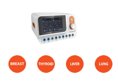
1. CuraWay US FDA RFA for Tumor, Hemorrhoid & Anal Fistula
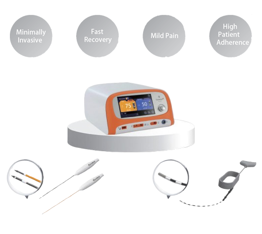
One of Unique Medi Trade's most innovative offerings is the CuraWay US FDA-Approved RFA (Radiofrequency Ablation) machine for the treatment of Tumor, Hemorrhoid and Anal Fistula. This machine represents the future of non-invasive treatments for these painful and often debilitating conditions.
-
Non-invasive treatment: The RFA machine uses high-frequency radio waves to target and shrink the problematic tissue, without the need for incisions.
-
Faster recovery: Compared to traditional surgical methods, RFA treatments result in quicker recovery times and less postoperative pain.
-
Minimally painful: Patients experience minimal discomfort during and after the procedure, making it a preferred choice for many.
The CuraWay RFA Machine is an example of how Unique Medi Trade has brought world-class medical technology to Bangladesh, offering healthcare professionals advanced tools to improve patient outcomes.
2. Handheld Wireless Color Doppler Ultrasound
The Handheld Wireless Color Doppler Ultrasound is another flagship product that exemplifies Unique Medi Trade’s commitment to making advanced medical technology more accessible. The device allows for high-quality, real-time imaging on a portable, easy-to-use platform, making it ideal for use in both clinical settings and remote areas.
Key features include:
-
Wireless and portable: The compact design allows for easy transportation and use in a variety of settings.
-
High-definition imaging: Provides clear, real-time images for accurate diagnostics.
-
Multi-functional: The device can be used for a range of applications, including vascular, abdominal, and obstetric imaging.
By offering such advanced, portable technology, Unique Medi Trade has greatly expanded the reach of ultrasound diagnostics across the country, particularly in rural and underserved regions.
3. ICU & OT Equipment
Unique Medi Trade also supplies ICU (Intensive Care Unit) and OT (Operating Theatre) equipment, ensuring hospitals have access to life-saving technology. From ventilators and defibrillators to surgical instruments, the company’s products help medical professionals provide the highest quality care to patients in critical conditions.
Unique Medi Trade’s Strengths
What makes Unique Medi Trade stand out in the crowded field of medical equipment suppliers? Here are some of the key strengths that have contributed to its leadership in the industry:
1. Exclusive Global Partnerships
Unique Medi Trade has established strategic partnerships with global medical equipment manufacturers, ensuring that it offers only the most reliable and innovative products available on the market. Notable collaborations include exclusive distribution rights for CuraWay, SonoHealth and SonoWave etc products in Bangladesh.
2. Comprehensive After-Sales Support
Unique Medi Trade offers unparalleled after-sales service, including:
-
Installation and training: Expert installation of medical equipment, followed by comprehensive training for medical staff to ensure proper use.
-
Maintenance and servicing: Routine maintenance, along with urgent repairs, ensures that equipment remains in optimal working condition.
-
Customer support: A dedicated support team available to assist clients with any issues or questions.
3. Fast Delivery and Logistics
Unique Medi Trade ensures that all orders are fulfilled in a timely manner, with fast and efficient delivery across the country. By partnering with reliable logistics providers, the company ensures that critical equipment reaches hospitals and clinics without delay, even in remote areas.
4. Innovation and Technology
Unique Medi Trade’s focus on introducing the latest medical technologies sets it apart from many competitors. The company continuously researches new developments in medical science and actively introduces the most innovative products to the Bangladeshi market.
Testimonials from Doctors and Hospitals
The trust that Unique Medi Trade has earned is evident in the positive feedback it receives from healthcare professionals. Surgeons, doctors, and hospital administrators across the country have shared their satisfaction with the company’s products and services. Here are a few examples of what they have to say:
-
Dr. Rajat Biswas, Associate Professor of Medicine, Chattagram Maa-O-Shishu Hospital: “Unique Medi Trade’s handheld ultrasound is a game-changer. Its portability and accuracy have greatly improved our diagnostic capabilities, especially in remote areas.”
-
Professor Dr. Md. Aminul Islam Joarder , BSMMU: “The CuraWay RFA machine has completely transformed the way we treat hemorrhoids and anal fistulas. It’s effective, fast, and causes minimal pain for our patients.”
Case Studies of Success
Unique Medi Trade has worked on several high-profile projects, providing medical equipment to major hospitals, diagnostic centers, and clinics across Bangladesh. Some notable installations include:
-
Dhaka Medical College Hospital: Installation of a range of diagnostic equipment, including Color Doppler Ultrasound, enhancing the hospital’s diagnostic capabilities.
-
Alliance Hospital: The adoption of RFA technology to treat patients with hemorrhoids and anal fistulas, resulting in faster recovery times and improved patient satisfaction.
Awards and Recognitions
Unique Medi Trade’s excellence in the medical equipment supply industry has not gone unnoticed. The company has been recognized with several prestigious awards for its innovation, customer service, and contribution to the healthcare sector.
3. SonoWave Technologies – Innovating for Tomorrow
Company Overview
SonoWave Technologies is a dynamic medical equipment supplier based in Bangladesh, renowned for its innovative approach to healthcare technology. Founded with the mission to bring cutting-edge diagnostic solutions to the healthcare sector, SonoWave has quickly risen to prominence in the Bangladeshi market.
The company's success lies in its ability to seamlessly blend advanced international technology with the needs of local healthcare providers. With a deep commitment to offering high-quality products and unparalleled customer support, SonoWave Technologies continues to shape the future of medical diagnostics in Bangladesh.
Core Values and Mission
SonoWave Technologies stands by its core values of integrity, innovation, and quality. The company’s mission is to provide healthcare providers in Bangladesh with access to the latest diagnostic tools, ensuring improved patient outcomes and more efficient care. The company firmly believes that technology should be both accessible and affordable, offering healthcare professionals the tools they need to save lives and enhance the quality of care.
Major Products and Solutions
SonoWave Technologies offers a diverse range of medical equipment, with a strong focus on ultrasound devices and diagnostic imaging solutions. These products cater to various healthcare needs, including obstetrics, gynecology, cardiology, and musculoskeletal imaging. Some of SonoWave's most prominent offerings include:
1. Portable Ultrasound Devices
The company's portable ultrasound devices are designed with the needs of busy healthcare professionals in mind. These devices combine portability with powerful imaging capabilities, allowing for accurate diagnosis even in remote or resource-limited areas. Features include:
-
Compact and lightweight: Easy to carry and store, making it ideal for use in field hospitals, rural clinics, or mobile medical units.
-
High-resolution imaging: Provides sharp, detailed images for accurate diagnoses, even for complex conditions.
-
User-friendly interface: Designed for ease of use, allowing healthcare providers to quickly learn the system and improve workflow efficiency.
SonoWave’s portable ultrasound devices are a significant step forward in ensuring that high-quality diagnostic imaging is available to everyone, regardless of location or infrastructure.
2. Color Doppler Ultrasound Systems
SonoWave’s Color Doppler Ultrasound systems offer high-definition imaging, providing real-time visualization of blood flow and tissue movement. These systems are indispensable in diagnosing vascular conditions, cardiovascular diseases, and fetal monitoring. Key features include:
-
Advanced color Doppler imaging: Enables the detection of blood flow abnormalities, such as clots, stenosis, or valve dysfunction.
-
Versatility: Used across multiple specialties, from cardiology to obstetrics, SonoWave’s Color Doppler systems are essential diagnostic tools in a wide array of clinical settings.
-
Portability: The Color Doppler devices from SonoWave are designed to be compact, making them suitable for both hospital use and smaller outpatient clinics.
3. Patient Monitoring Systems
In addition to diagnostic imaging, SonoWave Technologies also offers an array of patient monitoring systems. These systems are designed to continuously monitor critical patient parameters, ensuring timely interventions in emergencies. Products include:
-
Multi-parameter monitors: Track vital signs such as heart rate, blood pressure, temperature, and oxygen saturation.
-
Infusion pumps and ventilators: Essential for intensive care units (ICU) and emergency departments.
SonoWave’s patient monitoring solutions are built for accuracy and reliability, enabling healthcare teams to deliver real-time, data-driven care.
Competitive Edge
Several factors contribute to SonoWave Technologies' success and differentiate the company from other medical equipment suppliers in Bangladesh:
1. Technological Innovation
SonoWave stays ahead of the curve by continually introducing new and advanced technologies to the Bangladeshi market. Their products are sourced from renowned international manufacturers and are equipped with the latest features that allow healthcare providers to deliver high-quality care.
For example, SonoWave's portable ultrasound systems and color Doppler machines integrate AI and machine learning algorithms to assist doctors in analyzing imaging data, improving diagnostic accuracy, and reducing errors.
2. Affordable Pricing
While offering state-of-the-art technology, SonoWave Technologies has made it a point to offer its products at competitive prices. This affordability ensures that high-quality medical equipment remains accessible to a wider range of healthcare providers, including smaller clinics and rural hospitals.
3. Customer-Focused Service
Customer satisfaction is at the heart of SonoWave's operations. The company offers extensive post-sales support, including installation, training, and on-site servicing. Additionally, SonoWave’s dedicated customer service team ensures that healthcare providers can always access help when needed.
4. Comprehensive Warranty and Maintenance
SonoWave offers comprehensive warranties for all its products and ensures regular servicing and maintenance. This long-term support helps healthcare providers minimize downtime and maintain optimal device performance.
Partnerships and Distribution Channels
SonoWave Technologies has built strong relationships with international manufacturers, ensuring that its products are of the highest quality and meet international standards. The company’s exclusive partnerships with global brands enable it to distribute the latest and most reliable medical devices in Bangladesh.
SonoWave also collaborates with various hospitals, healthcare providers, and government organizations to ensure that its products reach the right institutions. This strong distribution network helps the company expand its footprint across the country, even in remote areas where quality medical equipment is hard to come by.
Notable Projects and Installations
SonoWave Technologies has been involved in numerous projects that have had a significant impact on Bangladesh’s healthcare landscape. Some key installations and projects include:
-
Installation of portable ultrasound systems in rural healthcare centers to improve maternal and fetal health monitoring in underserved areas.
-
Providing Color Doppler Ultrasound equipment to large private hospitals in Dhaka and Chittagong, enhancing their diagnostic capabilities in cardiology and vascular health.
-
Supply of patient monitoring systems to government hospitals across Bangladesh, helping improve critical care capabilities.
4: Praxis Health Care – Affordable & Quality Medical Equipment Provider
Company Overview
Praxis Health Care is a leading medical equipment supplier in Bangladesh, dedicated to offering high-quality yet affordable solutions to the country’s healthcare sector. Founded with the goal of meeting the growing demand for advanced medical equipment, Praxis Health Care has established itself as a trusted name in providing cutting-edge medical technology to hospitals, clinics, and healthcare facilities throughout Bangladesh.
What sets Praxis HealthCare apart is its focus on balancing the highest standards of technology with affordability. The company has managed to carve out a niche in the competitive medical supply market by catering to a diverse range of healthcare needs, from diagnostic imaging and patient monitoring to surgical instruments and laboratory equipment.
Core Values and Mission
Praxis Health Care’s mission revolves around three key pillars: affordability, quality, and reliability. The company is committed to delivering world-class medical equipment at prices that are accessible to both large hospitals and smaller healthcare providers, ensuring that healthcare professionals across Bangladesh have the tools they need to provide the best care for their patients.
The company’s core values of customer focus, integrity, and continuous improvement drive its operations. Praxis Health Care works relentlessly to maintain the trust of its customers by offering not only exceptional products but also outstanding after-sales support and service.
Major Products and Solutions
Praxis Health Care offers a diverse range of medical equipment, with a particular focus on diagnostic imaging, patient monitoring, and surgical instruments. Here are some of the company's major products and solutions:
1. Diagnostic Imaging Equipment
Praxis Health Care is renowned for its extensive selection of diagnostic imaging equipment, catering to various medical specialties. These devices provide high-resolution images to assist healthcare professionals in accurate diagnosis and treatment planning. Some of the key diagnostic imaging products offered by Praxis Health Care include:
-
Ultrasound Machines: Praxis provides a range of ultrasound machines that meet the needs of both small clinics and large hospitals. The devices are designed to provide clear, real-time imaging for a variety of applications, including obstetrics, gynecology, cardiology, and musculoskeletal imaging.
-
X-ray Machines: Offering both digital and analog X-ray machines, Praxis Health Care ensures that hospitals and clinics have access to advanced imaging technology at competitive prices.
-
CT Scanners: Praxis also supplies CT (Computed Tomography) scanners, essential for providing detailed cross-sectional images of the body, helping diagnose conditions such as tumors, internal bleeding, and bone fractures.
These diagnostic imaging devices are crucial for early detection, helping doctors make informed decisions about treatment plans and ultimately improving patient outcomes.
2. Patient Monitoring Systems
Patient monitoring is a critical aspect of healthcare, particularly in intensive care units (ICUs), emergency departments, and during surgery. Praxis Health Care’s patient monitoring systems provide real-time data to healthcare professionals, enabling them to respond quickly to changes in a patient’s condition. Key products include:
-
Multi-parameter Monitors: These monitors measure a variety of vital signs, including heart rate, blood pressure, oxygen saturation, and temperature. They are used to continuously track a patient’s status in both critical and non-critical settings.
-
ECG Machines: Electrocardiogram (ECG) machines offered by Praxis help monitor heart health by detecting abnormal heart rhythms and electrical activity.
-
Defibrillators: Praxis provides automated external defibrillators (AEDs), essential life-saving equipment used to restore normal heart rhythm in patients experiencing sudden cardiac arrest.
These systems are integral to ensuring that medical professionals have the necessary tools to provide timely interventions and manage critical patients.
3. Surgical Instruments and Equipment
Praxis Health Care is also a reliable supplier of high-quality surgical instruments and equipment. Whether it's for general surgery or specialized procedures, Praxis offers a comprehensive range of tools to support medical professionals during surgery. Some key offerings include:
-
Surgical Sets and Kits: Praxis supplies a wide range of surgical sets for various types of procedures, ensuring that healthcare providers have the necessary tools for efficient and safe surgeries.
-
Operating Tables: High-quality operating tables designed for various medical specialties, providing both comfort and stability for patients during surgery.
-
Surgical Lighting: LED surgical lights offering optimal illumination for precise operations. These lights reduce eye strain for surgeons and allow for better visibility of the surgical site.
These surgical tools and equipment contribute to the success of medical procedures and enhance the safety of both patients and surgeons.
Competitive Edge
Praxis Health Care’s success in the highly competitive medical equipment market in Bangladesh can be attributed to several key factors:
1. Affordability Without Compromising on Quality
Praxis Health Care stands out for offering affordable medical equipment without compromising on quality. This balance between cost and quality makes it accessible to a wide range of healthcare providers, from large hospitals to small clinics. By providing high-quality, cost-effective equipment, Praxis ensures that healthcare services are improved across all levels of the medical system.
2. Wide Product Range
Praxis Health Care offers a broad selection of products, ranging from diagnostic imaging tools to surgical instruments, making it a one-stop shop for healthcare professionals. This product diversity allows Praxis to cater to a wide array of medical specialties and ensures that its customers have access to everything they need for comprehensive patient care.
3. Customer Service and Support
One of the defining features of Praxis Health Care is its exceptional customer service. The company provides comprehensive after-sales support, including:
-
Installation and setup: Professional installation of medical equipment to ensure that devices are ready for immediate use.
-
Training: Praxis offers in-depth training programs for healthcare professionals to ensure that they are comfortable with the use of new equipment.
-
Maintenance and repair: The company offers regular maintenance services and repair support, ensuring that the equipment operates smoothly and without interruption.
These services are crucial in helping healthcare providers maximize the value of their investments and keep their operations running efficiently.
4. Partnerships with Renowned Manufacturers
Praxis Health Care works closely with some of the world’s leading medical equipment manufacturers, ensuring that it provides the latest technologies available on the market. These partnerships enable Praxis to bring international-quality products to Bangladesh at competitive prices, helping local healthcare professionals deliver world-class care.
Notable Projects and Installations
Praxis Health Care has been involved in numerous notable installations that have positively impacted the healthcare landscape in Bangladesh. Some of the key installations include:
-
Digital X-ray Systems: Installed in major hospitals across the country, Praxis’s digital X-ray systems have revolutionized the way radiology departments work, offering quicker and more accurate diagnoses.
-
ECG Machines: Installed in several cardiac care centers, these machines have helped doctors monitor and diagnose heart conditions effectively.
-
Operating Tables and Surgical Equipment: Praxis has provided hospitals with advanced operating tables and other essential surgical equipment, contributing to the successful outcomes of numerous surgeries.
5: SonoWave Medical System – A Global Leader in Medical Technology
Company Overview
SonoWave Medical System is a prominent player in the global medical equipment market, with a reputation for innovation, quality, and reliability. Based in China, SonoWave has become one of the leading manufacturers and suppliers of advanced medical equipment worldwide. The company specializes in ultrasound machines, color Doppler devices, and other diagnostic tools, offering cutting-edge solutions to healthcare providers across various regions, including Bangladesh.
Founded with the vision of improving healthcare through accessible and high-quality medical technology, SonoWave has garnered global recognition for its commitment to developing state-of-the-art devices. Its products are designed to improve diagnostic accuracy, patient care, and treatment outcomes while maintaining affordability for healthcare facilities in both developing and developed markets.
SonoWave Medical System is known for its robust research and development (R&D) department, which continuously works to bring new and advanced technologies to the market. The company also places a strong emphasis on customer satisfaction by providing timely support, service, and training to healthcare professionals around the world.
Core Values and Mission
At the heart of SonoWave’s operations are its core values of innovation, quality, and customer care. The company is driven by the desire to provide healthcare professionals with cutting-edge tools to deliver accurate diagnoses and effective treatments. SonoWave’s mission is to make advanced medical technology accessible, with a focus on improving global health outcomes.
The company’s commitment to continuous innovation means it is always at the forefront of medical technology, integrating the latest advancements to create high-performance devices. At the same time, its focus on quality control ensures that each product adheres to the highest international standards, making it a reliable choice for healthcare providers.
Major Products and Solutions
SonoWave Medical System offers a wide array of medical devices, but it is most renowned for its ultrasound machines, color Doppler devices, and portable diagnostic solutions. The company’s advanced medical equipment is used in a variety of specialties, including obstetrics, gynecology, cardiology, and musculoskeletal imaging. Let’s take a closer look at some of their flagship products:
1. Ultrasound Machines
SonoWave is widely recognized for its high-quality ultrasound machines that offer exceptional image clarity and diagnostic accuracy. These machines cater to a range of applications, from routine check-ups to complex medical imaging. Key features include:
-
High-resolution Imaging: SonoWave ultrasound machines provide detailed, high-resolution images that help healthcare professionals make accurate diagnoses.
-
Portable Models: The company offers portable ultrasound devices that allow healthcare providers to conduct on-site examinations, improving patient convenience and accessibility.
-
Advanced Doppler Ultrasound: With built-in Doppler imaging capabilities, SonoWave ultrasound machines are capable of providing real-time blood flow visualization, crucial for evaluating cardiac, vascular, and obstetric conditions.
These ultrasound machines are popular in both clinical and hospital settings for their versatility, ease of use, and ability to deliver fast, reliable results.
2. Color Doppler Ultrasound Systems
The Color Doppler ultrasound systems offered by SonoWave are particularly notable for their advanced capabilities in vascular and cardiac imaging. The technology allows for the assessment of blood flow within blood vessels, providing valuable information on a variety of medical conditions.
-
Comprehensive Imaging: SonoWave’s color Doppler systems provide a detailed, dynamic view of blood flow, allowing for the assessment of cardiovascular conditions such as arterial blockages and heart valve dysfunctions.
-
Real-time Visualization: These devices offer real-time color imaging, which is essential for accurate diagnosis and treatment planning in vascular, cardiac, and obstetric care.
-
Portable and Compact: Designed for both hospital and field use, SonoWave’s portable color Doppler systems combine high performance with portability, making them ideal for emergency rooms, outpatient clinics, and on-the-go applications.
Color Doppler systems from SonoWave have become an indispensable tool for cardiologists, obstetricians, and vascular surgeons.
3. Handheld and Portable Ultrasound Devices
One of the most groundbreaking products offered by SonoWave is its handheld and portable ultrasound devices. These compact and lightweight devices allow healthcare professionals to conduct imaging on the go, particularly in emergency and rural settings where access to full-sized ultrasound machines may be limited. Key benefits of SonoWave’s portable ultrasound systems include:
-
Convenience and Portability: Designed for easy transport and use in various settings, these handheld ultrasound devices are perfect for healthcare workers who need to perform diagnostics outside of a traditional hospital environment.
-
Ease of Use: SonoWave’s portable ultrasound devices are user-friendly and designed to be easily operated by healthcare professionals without extensive training.
-
Cost-Effective: These devices are an affordable alternative to traditional ultrasound machines, making diagnostic imaging more accessible to underserved populations and regions.
SonoWave’s handheld ultrasound devices have been instrumental in expanding healthcare access to remote areas, providing doctors and nurses with the ability to conduct critical diagnostic tests without the need for large, stationary equipment.
Competitive Edge
SonoWave Medical System has established itself as a leading name in the global medical equipment market due to several key factors that differentiate it from its competitors:
1. Innovative Technology
At the core of SonoWave’s competitive advantage is its focus on innovation. The company’s R&D team continuously works on developing new technologies that improve the functionality and performance of its products. This commitment to innovation has led to the creation of advanced imaging solutions, such as portable and handheld ultrasound systems, which provide healthcare professionals with greater flexibility and diagnostic capabilities.
2. Global Certifications and Standards
SonoWave Medical System holds multiple international certifications, including US FDA, CE, and ISO certifications, which ensure that their products meet global safety and quality standards. These certifications enhance the company’s reputation and provide healthcare providers with confidence in the reliability and effectiveness of their equipment.
3. Cost-Effectiveness
Despite offering cutting-edge medical equipment, SonoWave manages to keep its products affordable, making advanced medical technology accessible to a wide range of healthcare providers, particularly those in developing markets. The company’s commitment to delivering value for money has contributed to its rapid global expansion.
4. Excellent Customer Support
SonoWave’s dedication to customer satisfaction is evident through its robust customer service and support network. The company provides comprehensive training, installation, and maintenance services to healthcare professionals, ensuring that they can fully utilize their devices to improve patient care.
Notable Projects and Installations
SonoWave has been involved in numerous successful installations of its products in healthcare facilities across the globe, helping improve diagnostic capabilities and patient outcomes. Some of the most notable projects include:
-
Mobile Clinics in Rural Areas: SonoWave has supplied handheld ultrasound devices to mobile health clinics in rural and underserved areas, enabling healthcare providers to offer diagnostic imaging services to patients who would otherwise have limited access to medical care.
-
Hospitals and Diagnostic Centers: Many hospitals and diagnostic centers in Bangladesh and other countries have incorporated SonoWave’s ultrasound and color Doppler systems into their daily operations, improving diagnostic accuracy and patient satisfaction.
6: Conclusion and Future Outlook of the Medical Equipment Industry in Bangladesh
Conclusion
The medical equipment industry in Bangladesh has seen significant growth over the last decade, driven by the rising demand for healthcare services and technological advancements. Companies like Unique Medi Trade, SonoWave Technologies, Praxis Health Care, and SonoWave Medical System have played a pivotal role in bringing world-class medical devices to the country. These companies have not only introduced advanced medical technologies but also contributed to improving the quality of healthcare and accessibility in Bangladesh.
In this blog post, we have examined the leading medical equipment suppliers in Bangladesh, highlighting the key products and contributions of each company. From Unique Medi Trade's handheld wireless ultrasound devices to SonoWave’s portable ultrasound machines, these companies are ensuring that the healthcare industry in Bangladesh has access to the latest diagnostic and treatment tools.
The industry continues to evolve with the advent of new technologies and the expansion of healthcare infrastructure. As Bangladesh strives to meet the healthcare needs of its growing population, the role of medical equipment suppliers becomes increasingly important. They not only provide critical equipment but also offer essential services such as training, installation, and technical support to healthcare providers.
Key Takeaways:
-
Unique Medi Trade stands out as one of the premier suppliers of medical equipment in Bangladesh, offering advanced solutions such as handheld wireless ultrasound devices and RFA machines for hemorrhoids and anal fistulas.
-
SonoWave Technologies is a global leader in the medical equipment industry, known for its ultrasound machines and color Doppler systems. Their products cater to a wide range of medical specialties, including obstetrics, cardiology, and vascular imaging.
-
Praxis Health Care has earned its place as a trusted name in the industry, offering a diverse range of high-quality medical equipment that meets the needs of both local and international healthcare providers.
-
SonoWave Medical System, with its commitment to innovation and quality, has revolutionized the way healthcare professionals approach diagnostic imaging. Their portable and handheld ultrasound devices have greatly expanded access to healthcare in remote areas.
Each of these companies brings a unique set of products and services to the table, contributing to the development of Bangladesh's healthcare infrastructure. Their efforts align with the government's goal of achieving universal healthcare coverage and improving the standard of medical care for all citizens.
Future Outlook: Opportunities and Challenges
The future of the medical equipment industry in Bangladesh looks promising, with several opportunities on the horizon. However, there are also challenges that need to be addressed for continued growth and success. Let’s explore some of the key opportunities and challenges for the future of the medical equipment sector in Bangladesh.
1. Growing Demand for Healthcare Services
As the population of Bangladesh continues to grow, the demand for healthcare services is expected to increase significantly. This will drive the need for advanced medical equipment, particularly in areas such as diagnostic imaging, surgical tools, and critical care devices. The government’s focus on healthcare infrastructure development presents a significant opportunity for medical equipment suppliers to expand their presence and offerings.
2. Technological Advancements in Healthcare
The rapid pace of technological innovation in the medical field is a game-changer for the industry. From AI-driven diagnostics to robotic surgery, technological advancements are transforming healthcare delivery. Companies that are quick to adapt to these innovations will have a competitive edge. Bangladesh’s growing healthcare sector will likely benefit from cutting-edge medical equipment that incorporates the latest technologies, improving patient outcomes and operational efficiency in hospitals and clinics.
3. Increase in Medical Tourism
Bangladesh is gradually becoming a hub for medical tourism due to the availability of affordable healthcare services and treatments. As more international patients seek treatment in Bangladesh, the demand for high-quality medical equipment will rise. This opens up new opportunities for medical equipment suppliers to target both domestic and international markets, enhancing their global presence.
4. Improved Distribution Networks
One of the key factors driving the growth of the medical equipment industry is the improvement of distribution networks in Bangladesh. The increasing availability of high-quality products in both urban and rural areas is making advanced medical equipment more accessible to healthcare providers across the country. Companies are also focusing on providing training and support to ensure that healthcare professionals are well-equipped to operate advanced medical devices, further enhancing the effectiveness of the healthcare system.
5. Government Initiatives and Policies
The Bangladesh government is making significant investments in the healthcare sector through the development of new hospitals, clinics, and healthcare facilities across the country. The government’s commitment to universal health coverage and public health improvement provides a favorable environment for medical equipment suppliers to expand their offerings. Furthermore, policies that support local manufacturing and reduce import duties on medical devices can contribute to the growth of the industry.
6. **Challenges:
Despite the many opportunities, the medical equipment industry in Bangladesh faces several challenges:**
-
Regulatory Hurdles: The regulatory environment for medical equipment in Bangladesh can be complex, with stringent approval processes for certain products. Companies need to navigate these regulations to ensure that their products meet the required standards and are approved for use in the country.
-
Pricing and Affordability: While there is increasing demand for advanced medical devices, the affordability of these products remains a concern for many healthcare providers, particularly in rural areas. Companies need to find ways to offer cost-effective solutions without compromising on quality.
-
Healthcare Workforce Development: As medical technology becomes more sophisticated, there is a growing need for healthcare professionals to undergo continuous training. Ensuring that medical practitioners are properly trained to operate advanced equipment is crucial to maximizing the potential of these technologies.
7. The Role of Healthcare Financing
The expansion of healthcare financing options in Bangladesh, including medical insurance and affordable payment plans, will play a crucial role in increasing access to advanced medical equipment. Many healthcare providers, especially those in underserved areas, may struggle with the upfront costs of medical equipment. By introducing financing models, companies can help healthcare facilities manage costs while still obtaining the latest devices for their operations.
The Road Ahead for Medical Equipment Suppliers
The future of the medical equipment industry in Bangladesh is filled with both opportunities and challenges. Companies like Unique Medi Trade, SonoWave Technologies, Praxis Health Care, and SonoWave Medical System are poised to play an essential role in shaping the future of healthcare in the country. By continuing to innovate and improve their offerings, these companies can help meet the growing demand for medical equipment while addressing the needs of a diverse healthcare system.
Closing Thoughts
As the medical equipment industry in Bangladesh continues to grow, it will be important for suppliers to stay ahead of emerging trends and continuously improve their products and services. By fostering strong partnerships with healthcare providers, government institutions, and international organizations, medical equipment suppliers can help ensure that the country’s healthcare system remains capable of meeting the needs of its citizens.
The future of the medical equipment sector in Bangladesh is indeed promising, and the players within this market will continue to influence the nation’s health outcomes for years to come.
-


